作者投稿
作者投稿 专家审稿
专家审稿 编辑办公
编辑办公

Advances in Metrological Research of Optical Coherence Tomography in Ophthalmology
-
摘要: 从眼科OCT设备的原理、技术分类、商业发展、计量参数和标准器制作方面,讨论了眼科OCT的性能评价以及计量技术的发展。详细总结了眼科OCT设备的计量标准器、系统分辨率、信噪比/对比度、图像质量等计量评价手段方面的国内外研究进展,为眼科OCT技术标准化的进一步完善理清思路。Abstract: This paper discussed the performance evaluation of ophthalmic Optical Coherence Tomography (OCT) and the development of metrology technology in terms of the principles, technical classification, commercial development, metrology parameters, and standard production of ophthalmic OCT devices. This paper summarized the domestic and foreign research progress of measurement and evaluation methods such as measurement standard, system resolution, signal-to-noise ratio/contrast, and image quality of ophthalmic OCT equipment in detail, and clarified the ideas for the further improvement of ophthalmic OCT technology standardization.
-
Key words:
- optical coherence tomography /
- ophthalmology /
- retina /
- phantom /
- measurement /
- standard
-
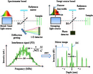
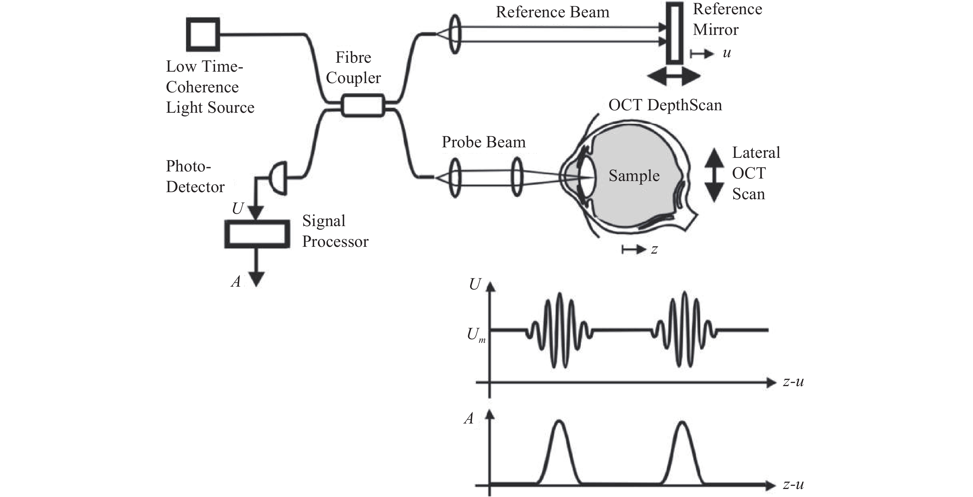
图 2 谱域OCT与扫频OCT技术原理
Figure 2. The principle of Spectral-Domain OCT and Swept-Source OCT
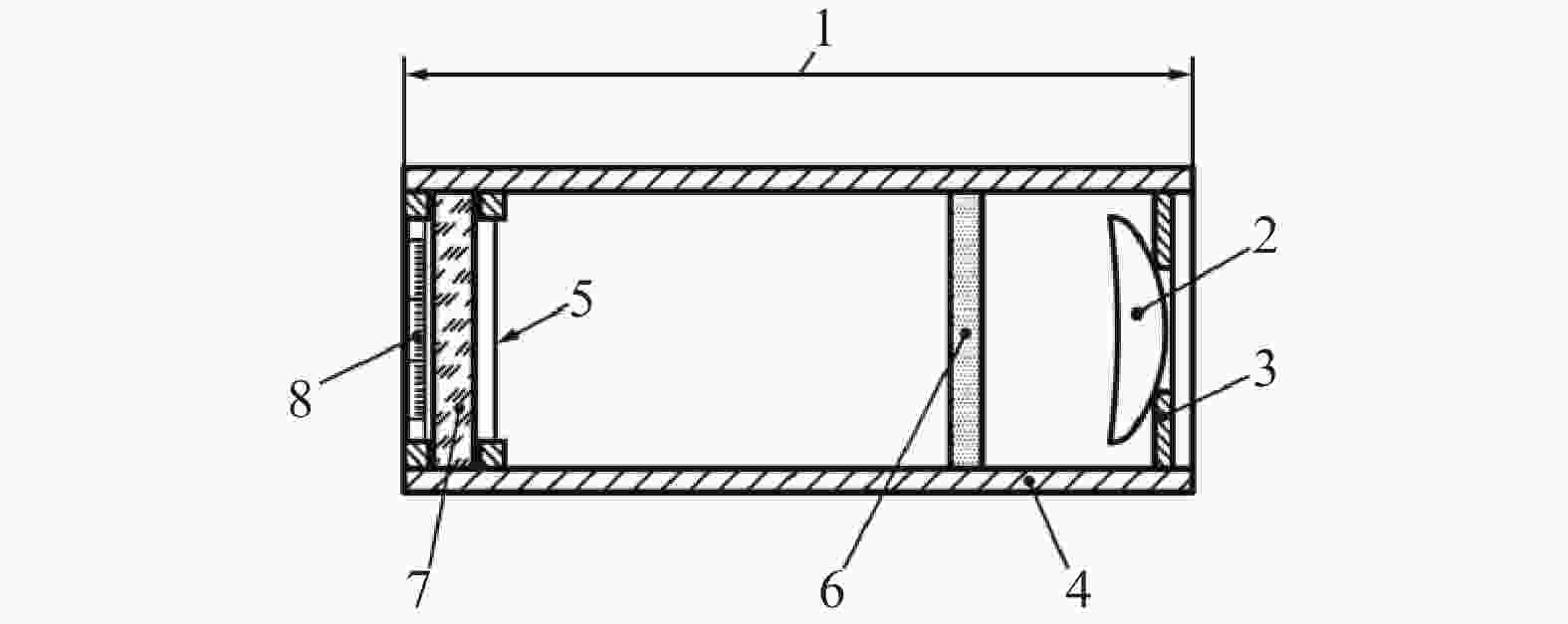
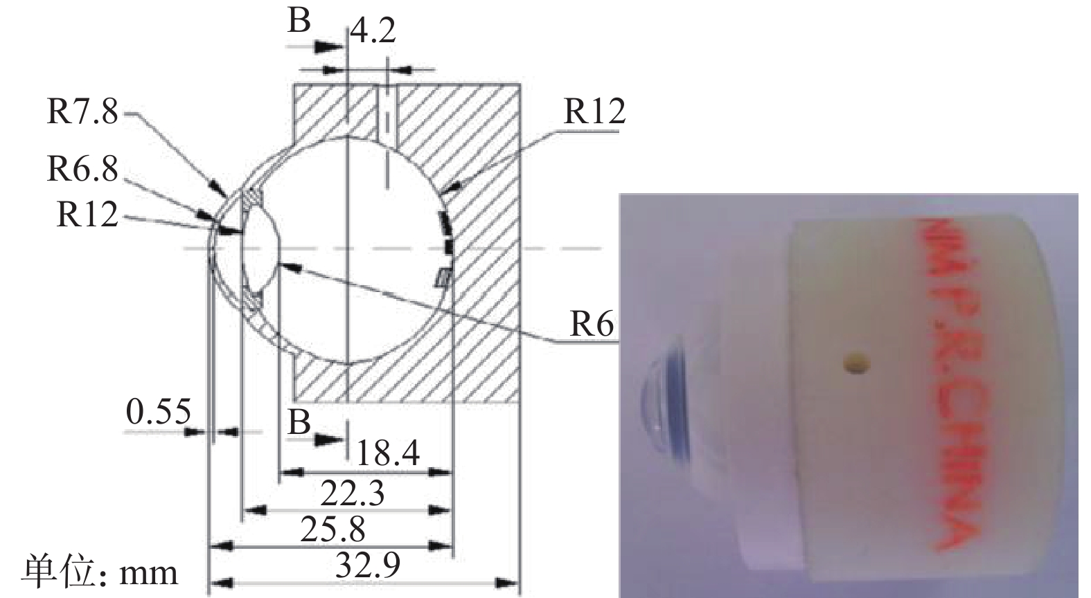
图 3 ISO 16971-2015推荐的计量标准器
1. 总长度17 mm;2. 镜头f= 17 mm;3. 孔径,直径6 mm;4. 管;5. 直径为100 µm的张紧细丝;6. 中性密度滤光片;7 .玻璃平面,厚度1 mm;8. 标尺
Figure 3. Reference standard recommended by ISO 16971-2015
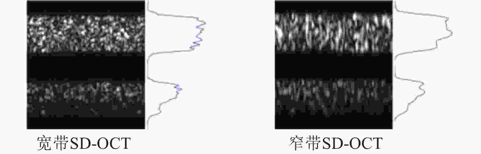
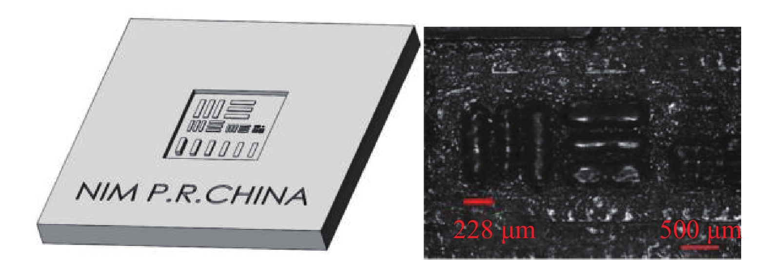
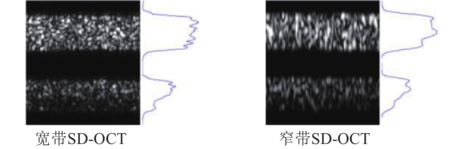
图 8 多层膜结构模体在系统中成像对比度差异
Figure 8. Imaging contrast differences of multilayer film structure phantom in in broadband systems
-
[1] FUJIMOTO J G, PITRIS C, BOPPART S A, et al. Optical Coherence Tomography: An Emerging Technology for Biomedical Imaging and Optical Biopsy[J]. Neoplasia, 2000, 2(1): 9-25. [2] BILLE J F. High Resolution Imaging in Microscopy and Ophthalmology: New Frontiers in Biomedical Optics[M]. Springer International Publishing, 2019. [3] HUANG D, SWANSON E A, LIN C P, et al. Optical Coherence Tomography[J]. Science, 1991, 254(5035): 1178-1181. doi: 10.1126/science.1957169 [4] FUJIMOTO J, SWANSON E. The Development, Commercialization, and Impact of Optical Coherence Tomography[J]. Investigative Ophthalmology & Visual Science, 2016, 57(9): 1-13. [5] FERCHER A F, HITZENBERGER C K, KAMP G, et al. Measurement of Intraocular Distances by Backscattering Spectral Interferometry[J]. Optics Communications, 1995, 117(1): 43-48. [6] CHINN S R, SWANSON E A, FUJIMOTO J G. Optical Coherence Tomography Using a Frequency-Tunable Optical Source[J]. Optics Letters, 1997, 22(5): 340-342. doi: 10.1364/OL.22.000340 [7] FERCHER A F, DREXLER W, HITZENBERGER C K. Optical Coherence Tomography - Principles and Applications[J]. Reports on Progress in Physics, 2003, 66(2): 239-244. doi: 10.1088/0034-4885/66/2/204 [8] DREXLER W, LIU M, KUMAR A, et al. Optical coherence tomography today: speed, contrast, and multimodality[J]. Journal of Biomedical Optics, 2014, 19(7): 071412. doi: 10.1117/1.JBO.19.7.071412 [9] REIF R, WANG R K. Optical Microangiography Based on Optical Coherence Tomography[M/OL]//Optical Coherence Tomography: Technology and Applications. Cham: Springer International Publishing, 2015: 1373–1397. [10] SHU X, BECKMANN L J, ZHANG H F. Visible-light optical coherence tomography: a review[J]. Journal of Biomedical Optics, 2017, 22(12): 121707. [11] KIRBY M A, PELIVANOV I, SONG S, et al. Optical coherence elastography in ophthalmology[J]. Journal of Biomedical Optics, 2017, 22(12): 121720. [12] BOER J F de, HITZENBERGER C K, YASUNO Y. Polarization Sensitive Optical Coherence Tomography – a Review [Invited][J]. Biomedical Optics Express, 2017, 8(3): 1838-1873. doi: 10.1364/BOE.8.001838 [13] JONNAL R S, KOCAOGLU O P, ZAWADZKI R J, et al. A Review of Adaptive Optics Optical Coherence Tomography: Technical Advances, Scientific Applications, and the Future[J]. Investigative Ophthalmology & Visual Science, 2016, 57(9): 51-68. [14] PIRCHER M, ZAWADZKI R J. Review of Adaptive Optics OCT (AO-OCT): Principles and Applications for Retinal Imaging [Invited][J]. Biomedical Optics Express, 2017, 8(5): 2536-2562. doi: 10.1364/BOE.8.002536 [15] KLEIN T, HUBER R. High-Speed OCT Light Sources and Systems [Invited][J]. Biomedical Optics Express, 2017, 8(2): 828-859. doi: 10.1364/BOE.8.000828 [16] KIM T S, JOO J, SHIN I, et al. 9.4 MHz A-Line Rate Optical Coherence Tomography at 1300 Nm Using a Wavelength-Swept Laser Based on Stretched-Pulse Active Mode-Locking[J]. Scientific Reports, 2020, 10(1): 9328-9335. doi: 10.1038/s41598-020-66322-0 [17] KOVACH J L, SCHWARTZ S G, FLYNN H W, et al. Anti-VEGF Treatment Strategies for Wet AMD[J]. Journal of Ophthalmology, 2012: 786870. [18] MINAKARAN N, DE CARVALHO E R, PETZOLD A, et al. Optical Coherence Tomography (OCT) in Neuro-Ophthalmology[J]. Eye, 2021, 35(1): 17-32. doi: 10.1038/s41433-020-01288-x [19] SHARMA D, AGRAWAL A, MATCHETTE L S, et al. Evaluation of a fiberoptic-based system for measurement of optical properties in highly attenuating turbid media.[J]. Biomedical engineering online, 2006, 5: 49-55. doi: 10.1186/1475-925X-5-49 [20] WANG Q, YANG H, AGRAWAL A, et al. Measurement of internal tissue optical properties at ultraviolet and visible wavelengths: Development and implementation of a fiberoptic-based system[J]. OPTICS EXPRESS, 2008, 16(12): 8685-8703. doi: 10.1364/OE.16.008685 [21] WANG Q, AGRAWAL A, WANG N S, et al. Evaluation of a reflectance-based approach for optical property determination in layered tissue[C]. Design and Quality for Biomedical Technologies II. SPIE, 2009: 105–115. [22] WANG Q, AGRAWAL A, WANG N S, et al. Condensed Monte Carlo Modeling of Reflectance From Biological Tissue With a Single Illumination-Detection Fiber[J]. IEEE JOURNAL OF SELECTED TOPICS IN QUANTUM ELECTRONICS, 2010, 16(3): 627-634. doi: 10.1109/JSTQE.2009.2029546 [23] ZAWADZKI R J, ROWE T S, FULLER A R, et al. Toward building an anatomically correct solid eye model with volumetric representation of retinal morphology[C]. Ophthalmic Technologies XX. SPIE, 2010: 412–418. [24] ROWE T S, ZAWADZKI R J. New developments in eye models with retina tissue phantoms for ophthalmic optical coherence tomography[C]. Optical Diagnostics and Sensing XII: Toward Point-of-Care Diagnostics; and Design and Performance Validation of Phantoms Used in Conjunction with Optical Measurement of Tissue IV. SPIE, 2012: 208–215. [25] ROWE T S, ZAWADZKI R J. Development of a corneal tissue phantom for anterior chamber optical coherence tomography (AC-OCT)[C]. Design and Performance Validation of Phantoms Used in Conjunction with Optical Measurement of Tissue V. SPIE, 2013: 84–92. [26] AGRAWAL A, BAXI J, CALHOUN W, et al. Optic Nerve Head Measurements With Optical Coherence Tomography: A Phantom-Based Study Reveals Differences Among Clinical Devices[J]. INVESTIGATIVE OPHTHALMOLOGY & VISUAL SCIENCE, 2016, 57(9): 413-420. [27] LEE H-J, LEE H-J, SAMIUDIN N M, et al. Retina Phantom for the Evaluation of Optical Coherence Tomography Angiography Based on Microfluidic Channels[J]. Biomedical Optics Express, 2019, 10(11): 5535-5548. doi: 10.1364/BOE.10.005535 [28] PFEFER J, AGRAWAL A. A review of consensus test methods for established medical imaging modalities and their implications for optical coherence tomography[C]. Design and Quality for Biomedical Technologies V. SPIE, 2012: 65–74. [29] 胡志雄, 郝冰涛, 孙欣, 等. 眼科光学相干断层(OCT)成像设备的计量研究[J]. 中国计量, 2016(7): 80-82. [30] 胡志雄, 刘文丽, 洪宝玉, 等. 光学相干层析成像三维分辨率测试模拟眼[J]. 光电工程, 2014, 41(12): 28-32, 38. [31] 胡志雄, 郝冰涛, 刘文丽, 等. 用于光学相干层析成像设备点扩散函数测量的模体制作与使用方法研究[J]. 光学学报, 2015, 35(4): 283-289. [32] CAO Z, DING Z, HU Z, et al. A standard model eye with micro scale multilayer structure for ophthalmic optical coherence tomography equipment[C]. Optical Measurement Technology and Instrumentation. SPIE, 2016: 626–632. [33] TOMLINS P H, FERGUSON R A, HART C, et al. Point-Spread Function Phantoms for Optical Coherence Tomography. [EB/OL](2009-8-1).https://eprintspublications.npl.co.uk/4463/. [34] AGRAWAL A, PFEFER T J, GILANI N, et al. Three-Dimensional Characterization of Optical Coherence Tomography Point Spread Functions with a Nanoparticle-Embedded Phantom[J]. Optics Letters, 2010, 35(13): 2269-2271. doi: 10.1364/OL.35.002269 [35] AGRAWAL A, CHANG R, CONNORS M, et al. System-independent assessment of OCT axial resolution with a "bar chart" phantom[J]. Optical Engineering, 2011, 7906: 880958. [36] AGRAWAL A, CONNORS M, BEYLIN A, et al. Characterizing the point spread function of retinal OCT devices with a model eye-based phantom[J]. BIOMEDICAL OPTICS EXPRESS, 2012, 3(5): 1116-1126. doi: 10.1364/BOE.3.001116 [37] FOUAD A, PFEFER T J, CHEN C-W, et al. Variations in optical coherence tomography resolution and uniformity: a multi-system performance comparison[J]. BIOMEDICAL OPTICS EXPRESS, 2014, 5(7): 2066-2081. doi: 10.1364/BOE.5.002066 [38] KEDIA N, LIU Z, SOCHOL R D, et al. 3-D printed photoreceptor phantoms for evaluating lateral resolution of adaptive optics imaging systems[J]. OPTICS LETTERS, 2019, 44(7): 1825-1828. doi: 10.1364/OL.44.001825 [39] LAMONT A C, RESTAINO M A, ALSHARHAN A T, et al. Direct laser writing of a titanium dioxide-laden retinal cone phantom for adaptive optics-optical coherence tomography[J]. OPTICAL MATERIALS EXPRESS, 2020, 10(11): 2749-2759. doi: 10.1364/OME.396150 [40] HU Z, HAO B, LIU W, et al. Test target for characterizing 3D resolution of optical coherence tomography[C]. International Symposium on Optoelectronic Technology and Application 2014: Laser and Optical Measurement Technology; and Fiber Optic Sensors. SPIE, 2014: 398–404. [41] FU X, HU Z, GE C, et al. A miniaturized and integrated system to measure key parameters of ophthamic optical coherence tomography equipment[C]. 2015 International Conference on Optical Instruments and Technology: Optoelectronic Measurement Technology and Systems. SPIE, 2015: 124–132. [42] 付晓宇, 胡志雄, 葛春风, 等. 眼科光学相干层析成像设备分辨率关键参数的小型化检测装置研制[J]. 计量学报, 2017, 38(6): 690-692. doi: 10.3969/j.issn.1000-1158.2017.06.07 [43] WEN T, DONG J, HU Z, et al. A standard test method based on point spread function for three-dimensional imaging system[C]. 8th International Symposium on Advanced Optical Manufacturing and Testing Technologies: Optical Test, Measurement Technology, and Equipment. SPIE, 2016: 608–616. [44] CAO Z, DING Z, HU Z, et al. Model eyes with curved multilayer structure for the axial resolution evaluation of an ophthalmic optical coherence tomography device[J]. Journal of Innovative Optical Health Sciences, 2018, 11(3): 1850013. doi: 10.1142/S179354581850013X [45] HUANG N, DENG Z, HU Z, et al. A Spatial Resolution Evaluation Method of Endoscopic Optical Coherence Tomography System Using the Annular Phantom[J]. Journal of Biophotonics, 2021, 14(8): 202100035. [46] AGRAWAL A, HUANG S, WEI HAW LIN A, et al. Quantitative evaluation of optical coherence tomography signal enhancement with gold nanoshells.[J]. Journal of biomedical optics, 2006, 11(4): 041121. doi: 10.1117/1.2339071 [47] Heikka T, Ometto G, Rowe T, et al. Testing a Phantom Eye under Various Signal-to-Noise Ratio Conditions Using Eleven Different OCT Devices[J]. Biomedical Optics Express, 2020, 11(3): 1306-1315. doi: 10.1364/BOE.383103 [48] AGRAWAL A, CHEN C-W, BAXI J, et al. Multilayer thin-film phantoms for axial contrast transfer function measurement in optical coherence tomography[J]. BIOMEDICAL OPTICS EXPRESS, 2013, 4(7): 1166-1175. doi: 10.1364/BOE.4.001166 [49] WOOLSEY N, WANG H-W, AGRAWAL A, et al. Quantitative analysis of low contrast detectability in optical coherence tomography[C]. Smart Biomedical and Physiological Sensor Technology XI. SPIE, 2014: 40–47. [50] AGRAWAL A, PFEFER T J, WOOLLIAMS P D, et al. Methods to assess sensitivity of optical coherence tomography systems[J]. BIOMEDICAL OPTICS EXPRESS, 2017, 8(2): 902-917. doi: 10.1364/BOE.8.000902 [51] BAXI J, CALHOUN W, SEPAH Y J, et al. Retina-simulating phantom for optical coherence tomography[J]. JOURNAL OF BIOMEDICAL OPTICS, 2014, 19(2): 1106-1114. [52] LOZZI A, AGRAWAL A, BORETSKY A, et al. Image quality metrics for optical coherence angiography[J]. BIOMEDICAL OPTICS EXPRESS, 2015, 6(7): 2435-2447. doi: 10.1364/BOE.6.002435 [53] 李修宇, 吴福宝, 胡志雄, 等. 眼科光学相干断层扫描成像设备关键参数计量技术研究[J]. 中国医疗设备, 2019, 34(11): 16-21, 29. doi: 10.3969/j.issn.1674-1633.2019.11.004 [54] WANG H, LIU W, HU Z, et al. Model eye tool for retinal optical coherence tomography instrument calibration[J]. Journal of Innovative Optical Health Sciences, 2021, 14(3): 2150010. doi: 10.1142/S1793545821500103 -

 下载:
下载:


 点击查看大图
计量
- 文章访问数: 722
- HTML全文浏览量: 227
- PDF下载量: 93
- 被引次数: 0