 作者投稿
作者投稿 专家审稿
专家审稿 编辑办公
编辑办公

Study on the Impact of Cell Viability Detection and Processing on Drug Effect Evaluation
-
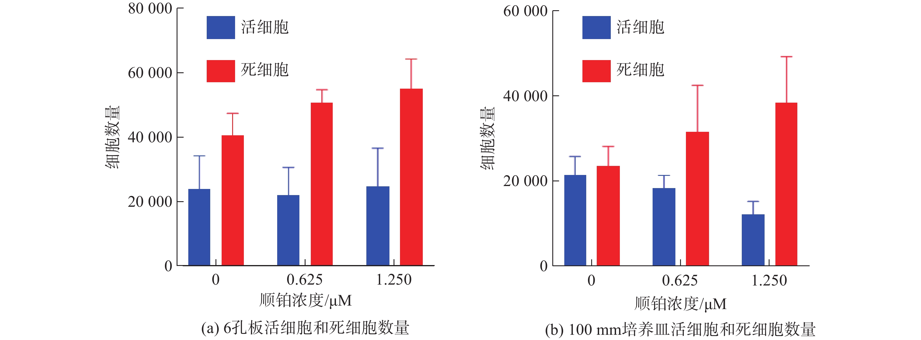
摘要: 在使用体外培养的细胞进行药物实验时,药物处理会造成细胞死亡,而检测细胞存活率的方法不同,会对实验结果造成影响。以海拉细胞作为实验对象,用不同浓度的顺铂处理细胞,做去除以及不去除旧培养基的处理,比较包含或不包含旧培养基中漂浮细胞情况下检测得到的细胞存活率。实验结果显示,使用CCK8法检测96孔板、24孔板细胞存活率,不去除旧培养基时实验组细胞存活率高于去除旧培养基实验组细胞存活率。在顺铂浓度为0.625 μM时,不去除旧培养基与去除旧培养基实验组之间的细胞存活率有显著差异。在6孔板和100 mm培养皿中,使用细胞计数仪对用药后的细胞培养基中的漂浮细胞进行检测,发现培养基中漂浮的细胞中存在活细胞。结果表明药物处理后的培养基中存在漂浮的活细胞,去除旧培养基会去除这部分活细胞,从而影响细胞药物实验结果的准确性。Abstract: When conducting drug experiments on cells cultured in vitro, drug treatment can cause cell death, and different methods for detecting cell survival rates can have impacts on the experimental results. This experiment takes Hela cells as the experimental object, and treats the cells with different concentrations of cisplatin. The cell survival rates detected with or without floating cells in the old culture medium are compared. The experimental results indicated that using the CCK8 method to detect the cell survival rates of 96 well plates and 24 well plates, the cell survival rates of the experimental group without removing the old culture medium was higher than that of the experimental group removing the old culture medium. At a cisplatin concentration of 0.625 μM, there is a significant difference in cell survival rates between the experimental group with and without removing the old medium. In 6-well plates and 100 mm dishes, a cell counter was used to detect the floating cells in the cell culture medium after application of drugs, and the presence of viable cells in the floating cells in the medium was found. Therefore, the floating living cells in the culture medium will affect the experimental results of cell drug experiment.
-
Key words:
- metrology /
- cell cytometry /
- detection method /
- cell viability /
- drug effects /
- in vitro culture
-
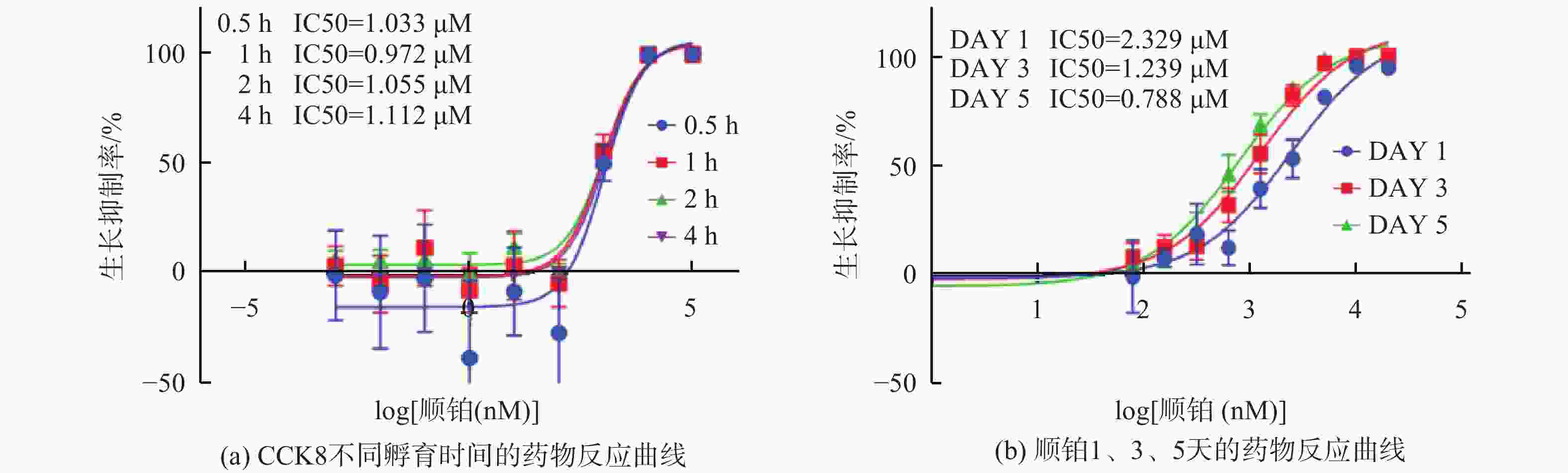
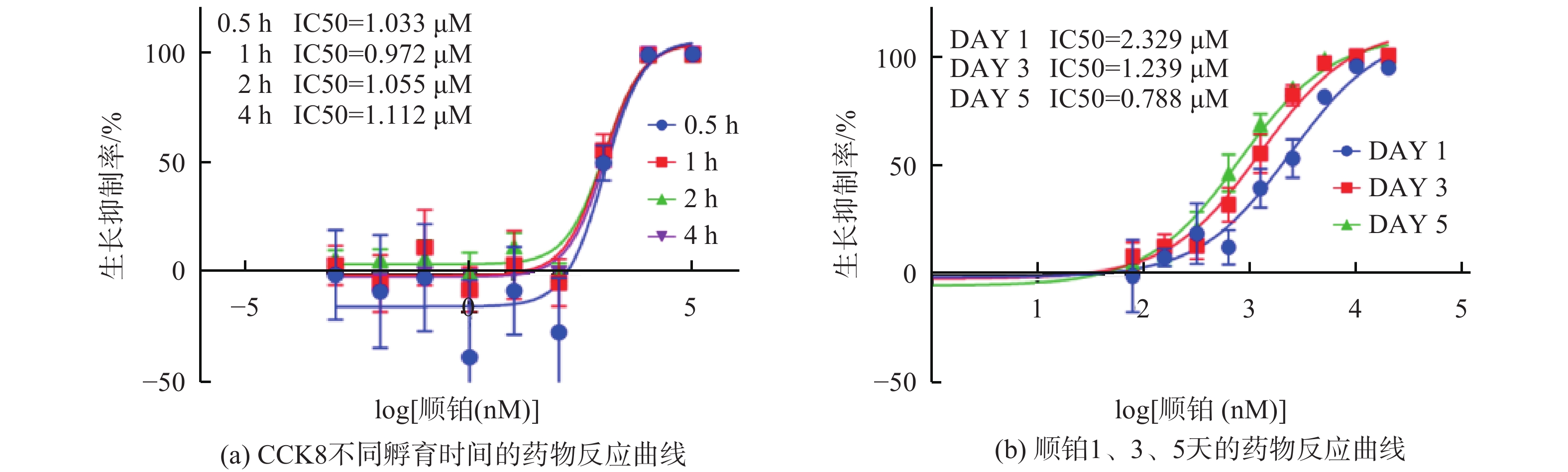
图 1 不同药物浓度和不同CCK8孵育时间下的药物作用
Figure 1. The drug effect under different drug concentrations and different CCK8 incubation time
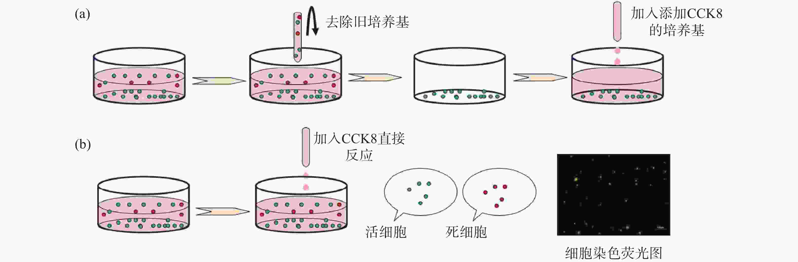
图 2 培养基不同处理方式操作示意图
Figure 2. Schematic diagram of the operation of different processing methods of culture medium
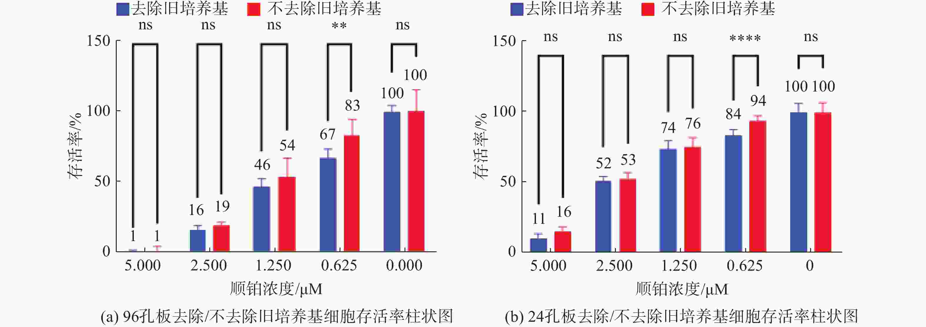
图 3 去除/不去除旧培养基检测的细胞存活率
Figure 3. Cell viability assays with or without removal of old media
-
[1] Lian-Shun Feng. Development and Advances of Drugs for Cancer Theranostics – PART-IV[J]. Current Topics in Medicinal Chemistry, 2021, 18(21): 1644. [2] Tosca E. M, Ronchi D, Facciolo D, et al. Replacement, Reduction and Refinement of Animal Experiments in Anticancer Drug Development: The Contribution of 3D In Vitro Cancer Models in the Drug Efficacy Assessment[J]. Biomedicines, 2023, 11: 1058. doi: 10.3390/biomedicines11041058 [3] 薛志超, 赵佳威, 李永淑, 等. 细胞密度对抗癌药药效评估准确性的研究[J]. 计量科学与技术, 2023, 67(4): 57-62,56. [4] Jaroch K, Jaroch A, Bojko B. Cell cultures in drug discovery and development: The need of reliable in vitro-in vivo extrapolation for pharmacodynamics and pharmacokinetics assessment[J]. Journal of Pharmaceutical and Biomedical Analysis, 2018, 147: 297-312. doi: 10.1016/j.jpba.2017.07.023 [5] Schilling K, Harris AL, Halliday AN, et al. Investigations on Zinc Isotope Fractionation in Breast Cancer Tissue Using in vitro Cell Culture Uptake-Efflux Experiments[J]. Front. Med, 2022, 8: 746532. doi: 10.3389/fmed.2021.746532 [6] Faruqui Nilofar, Kummrow Andreas, Fu Boqiang, et al. Cellular Metrology: Scoping for a Value Proposition in Extra- and Intracellular Measurements[J]. Frontiers in Bioengineering and Biotechnology, 2020, 7: 456. doi: 10.3389/fbioe.2019.00456 [7] Mirabelli, Coppola, Salvatore. Cancer Cell Lines Are Useful Model Systems for Medical Research[J]. Cancers, 2019, 11(8): 1098. doi: 10.3390/cancers11081098 [8] 薛志超, 曾嘉明, 李永淑, 等. 接种数量对细胞生长和药物作用的影响及细胞计数方法对比[J]. 计量学报, 2023, 3(44): 341-349. [9] Minerva, Amrita Bhat, Sonali Verma, et al. Cisplatin-based combination therapy for cancer[J]. Journal of Cancer Research and Therapeutics, 2023, 19(3): 530-536. doi: 10.4103/jcrt.jcrt_792_22 [10] Raudenska Martin, Balvan Jan, Fojtu Michaela, et al. Unexpected therapeutic effects of cisplatin.[J]. Metallomics, 2019, 11(7): 1182-1199. doi: 10.1039/c9mt00049f [11] Mengdi Song, Mingxiao Cui, Kehai Liu. Therapeutic strategies to overcome cisplatin resistance in ovarian cancer[J]. European Journal of Medicinal Chemistry, 2022, 232: 114205. doi: 10.1016/j.ejmech.2022.114205 [12] Chenying Jiang, Chenjun Shen, Maowei Ni, et al. Molecular mechanisms of cisplatin resistance in ovarian cancer[J]. Genes & Diseases, 2023, 1: 1. [13] Liang C, Zhang Hy, Wang Yq, et al. TMED2 Induces Cisplatin Resistance in Breast Cancer via Targeting the KEAP1-Nrf2 Pathway[J]. CURR MED SCI, 2023, 43: 1023-1032. doi: 10.1007/s11596-023-2777-7 [14] alma Y Mohamed, Hisham A Elshoky, Nayera M El-Sayed, et al. Ameliorative effect of zinc oxide-chitosan conjugates on the anticancer activity of cisplatin: Approach for breast cancer treatment[J]. International Journal of Biological Macromolecules, 2023, 257: 128597. [15] A Rane, J Jarmoshti, A Siddique, et al. Dielectrophoretic enrichment of live chemo-resistant circulating-like pancreatic cancer cells from media of drug-treated adherent cultures of solid tumors[J]. Lab Chip, 2023, 6: 10. [16] Grela E, Kozłowska J, Grabowiecka A. Current methodology of MTT assay in bacteria – A review[J]. Acta Histochemica, 2018, 120(4): 303-311. doi: 10.1016/j.acthis.2018.03.007 [17] Ludmil Benov. Improved Formazan Dissolution for Bacterial MTT Assay[J]. Microbiology Spectrum, 2021, 9(3): 1. [18] Yanfeng Zhu, Weihui Chen, Weiqun Guan, et al. Study of As2O3 regulating proliferation and apoptosis of Tca8113 cells by inhibiting the expression of Id-1, Artificial Cells[J]. Nanomedicine, and Biotechnology, 2019, 47(1): 1932-1937. [19] Jun Wang , Wei Lei, Gang Li, et al. CD151 promotes proliferation and migration of SK-NEP-1 cells via the GSK-3β/P21/cyclinD signaling pathway[J]. Pathlogy-Research and Practice, 2019, 215(2): 329-334. [20] Ling Cai, Xijiang Qin, Zhihui Xu, et al. Comparison of Cytotoxicity Evaluation of Anticancer Drugs between Real-Time Cell Analysis and CCK-8 Method[J]. ACS Omega, 2019, 4: 12036-12042. doi: 10.1021/acsomega.9b01142 [21] Jing-Ru Song, Na Li, Dian-Peng Li. Synthesis and anti-proliferation activity of mogrol derivatives bearing quinoline and triazole moieties[J]. Bioorganic & amp; Medicinal Chemistry Letters, 2021, 42: 128090. [22] de la Fuente-Jiménez J L, Rodríguez-Rivas C I, Mitre-Aguilar I B, et al. A Comparative and Critical Analysis for In Vitro Cytotoxic Evaluation of Magneto-Crystalline Zinc Ferrite Nanoparticles Using MTT, Crystal Violet, LDH, and Apoptosis Assay[J]. Mol. Sci, 2023, 24: 12860. doi: 10.3390/ijms241612860 [23] Magdalena Boncler, Marek Różalski, Urszula Krajewska, et al. Comparison of PrestoBlue and MTT assays of cellular viability in the assessment of anti-proliferative effects of plant extracts on human endothe[J]. Journal of Pharmacological and Toxicological Methodslial cells, 2014, 69(1): 9-16. doi: 10.1016/j.vascn.2013.09.003 [24] Kumar Priti, Nagarajan Arvindhan, Uchil Pradeep D. Analysis of Cell Viability by the MTT Assay[J]. Cold Spring Harbor Protocols, 2018, 6: 469-472. [25] Yu C, Zhang X, Wang M, et al. Afatinib combined with anti-PD1 enhances immunotherapy of hepatocellular carcinoma via ERBB2/ STAT3/PD-L1 signaling[J]. Front Oncol, 2023, 13: 1198118. doi: 10.3389/fonc.2023.1198118 [26] Wang Junbin, Gao Jin, Chen Qinnan, et al. LncRNA LINC01116 Contributes to Cisplatin Resistance in Lung Adenocarcinoma[J]. OncoTargets and Therapy, 2020, 13: 9333-9348. doi: 10.2147/OTT.S244879 [27] anfang Wang, Zhenhao Zhang, Wei Wan, et al. FAM19A5/S1PR1 signaling pathway regulates the viability and proliferation of mantle cell lymphoma[J]. Journal of Receptors and Signal Transduction, 2022, 42(3): 225-229. doi: 10.1080/10799893.2021.1895220 [28] Wang Ya, Zhang Xiaomei, Zhao Boyuan, et al. Suspension State Promotes Drug Resistance of Breast Tumor Cells by Inducing ABCC3 Overexpression[J]. Applied Biochemistry and Biotechnology, 2020, 190: 410-422. doi: 10.1007/s12010-019-03084-0 [29] Mao Yuqiang, Yu Ying, Han Yun. Influence of thoracic drainage fluid on proliferation, migration, apoptosis, and drug resistance in lung cancer cell lines[J]. Cancer Management and Research, 2019, 11: 2253-2259. doi: 10.2147/CMAR.S187019 [30] Zhuo-Ya DAI, Shuang-Mei JIN, Hong-Qin LUO, et al. LncRNA HOTAIR regulates anoikis-resistance capacity and spheroid formation of ovarian cancer cells by recruiting EZH2 and influencing H3K27 methylation[J]. Neoplasma, 2021, 68(3): 509-518. doi: 10.4149/neo_2021_201112N1212 [31] Grimes D R, Fletcher A G. Close Encounters of the Cell Kind: The Impact of Contact Inhibition on Tumour Growth and Cancer Models[J]. Bulletin of Mathematical Biology, 2020, 82(2): 20. doi: 10.1007/s11538-019-00677-y -

 下载:
下载:


 点击查看大图
图(5)
计量
- 文章访问数: 57
- HTML全文浏览量: 58
- PDF下载量: 0
- 被引次数: 0

