 作者投稿
作者投稿 专家审稿
专家审稿 编辑办公
编辑办公

Overview of the Development of Near-Field Optical Microscopy
-
摘要: 半导体芯片制造、超精密加工以及生物医学等领域的探索已经深入纳米尺度,对相应的测量技术提出了更高要求。光学方法以其非接触、无损、快速等优势得到了广泛应用,但传统光学显微镜的分辨率受限于衍射极限。扫描近场光学显微镜基于非辐射场的探测和成像原理,能够突破衍射极限,在超高光学分辨率下进行纳米尺度光学成像。介绍了近场光学显微镜的成像原理,分析了其不同工作方式下的优缺点。针对目前近场光学显微镜发展过程中所聚焦的提高分辨率和信噪比两个重点方向进行了综述。在提高分辨率方面,通过在孔径式近场光学显微镜探针尖端添加纳米天线,基于纳米天线原理突破了探针孔径对于分辨率的限制;利用在光纤尖端包覆金属膜或嵌入金属纳米线等方法,将光纤内的光转化为表面等离激元形式传播至尖端实现高分辨测量;利用一种探针与样品的极小间距模式将光斑压缩,突破散射式近场光学显微镜分辨率受针尖大小的限制,提高了分辨率。在增强针尖处聚焦光场强度、提高信噪比研究方面,通过在探针尖端制备各种纳米结构来激发表面等离激元,避免了远场光直射针尖区域所造成的背景噪声,提高了信噪比;在光纤式探针中通过增加环形光栅等结构来提高表面等离激元转化效率,增加聚焦光场强度,提高了信号强度;采用平台等特殊结构使表面等离激元产生反射共振增强,提高聚焦光场强度。最后,总结并展望了未来近场光学显微镜的发展方向。Abstract: As the semiconductor industry, ultra-precision machining, and biomedical fields delve into the nanoscale, the demand for advanced measurement technologies has heightened. Optical methods, with their non-contact, non-destructive, and rapid properties, have been widely employed, yet the resolution of traditional optical microscopes is bounded by the diffraction limit. Scanning Near-Field Optical Microscopy (SNOM), operating on the principles of non-radiative field detection and imaging, overcomes this limit to facilitate nanoscale optical imaging with ultra-high resolution. This review introduces the imaging principles of near-field optical microscopy and analyzes its advantages and disadvantages across various operational modes. It then delves into two crucial aspects of SNOM's ongoing development: enhancing resolution and signal-to-noise ratio (SNR). In terms of improving resolution, approaches like adding nano-antennas to the tips of aperture-type SNOM probes have been explored to break through the probe aperture's resolution constraint. Techniques such as coating optical fibers with metal films or embedding metal nanowires to convert light into surface plasmon forms for high-resolution measurements are discussed. Additionally, the use of a minimal gap mode between the probe and sample to compress the light spot, thereby exceeding the resolution limitations of scattering-type SNOMs, is examined. The review also addresses strategies to augment the light field intensity at the probe tip and improve SNR. These include fabricating various nanostructures at the probe tip to excite surface plasmons, thereby mitigating background noise caused by direct far-field light, and enhancing the SNR. Innovations in fiber-optic probes, such as the inclusion of ring gratings to improve surface plasmon conversion efficiency, are explored to increase the focused light field's intensity and signal strength. The utilization of special structures like platforms to induce surface plasmon reflective resonance enhancement, thus increasing the focused light field's intensity, is also discussed. The article concludes by summarizing the state of near-field optical microscopy and projecting its future development trajectory.
-
Key words:
- metrology /
- near-field optical microscopy /
- resolution /
- signal-to-noise ratio /
- surface plasmon /
- nano-focusing
-
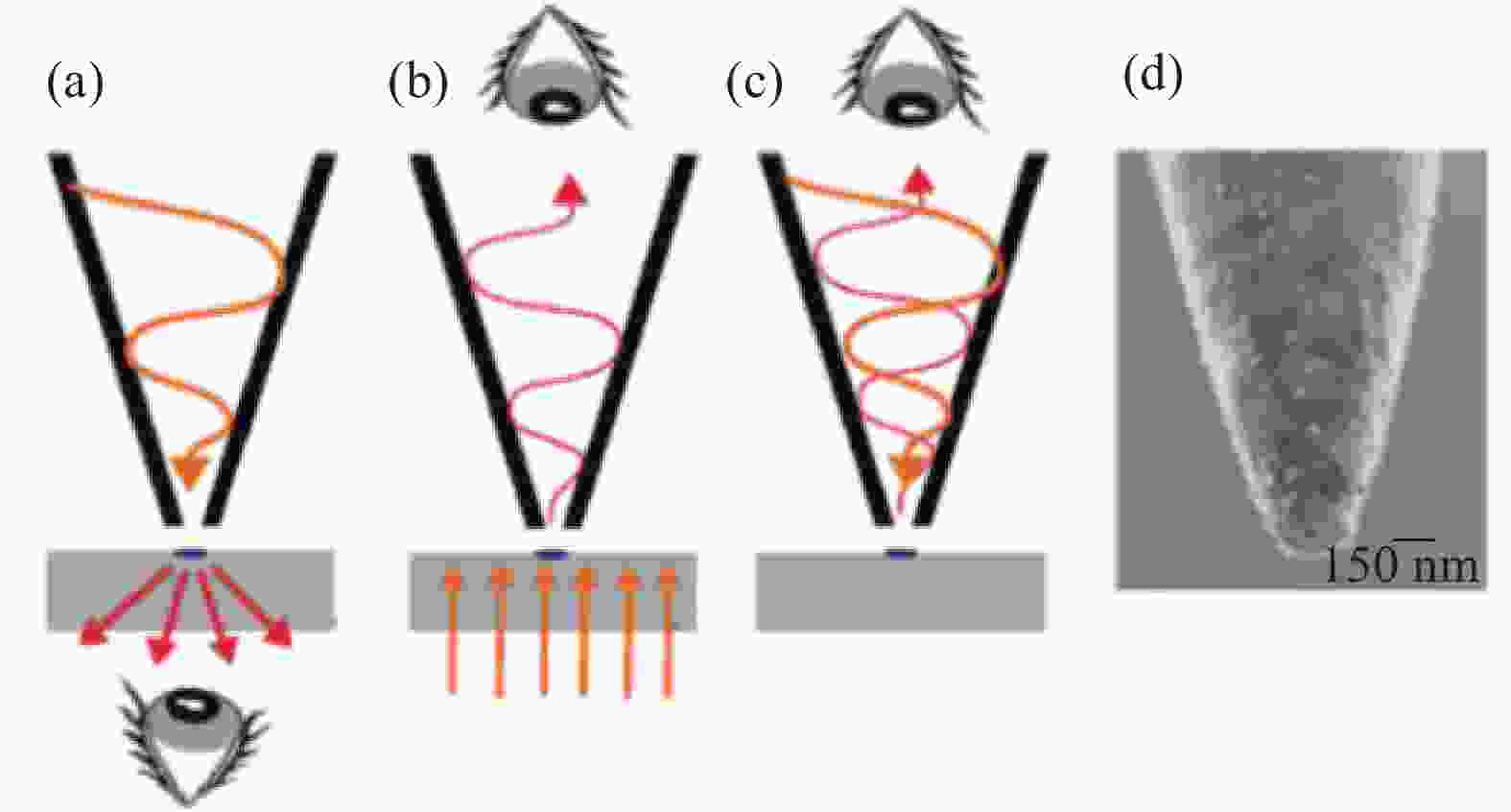
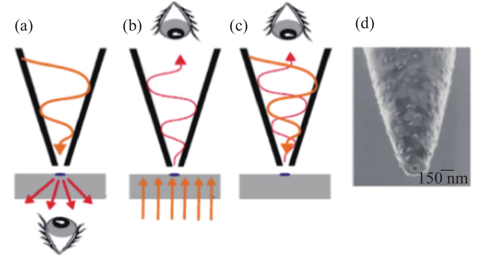
图 3 A-SNOM不同工作模式
注:a)照明模式;b)收集模式;c)照明-收集模式;d)有孔探针电镜图。
Figure 3. A-SNOM different working modes

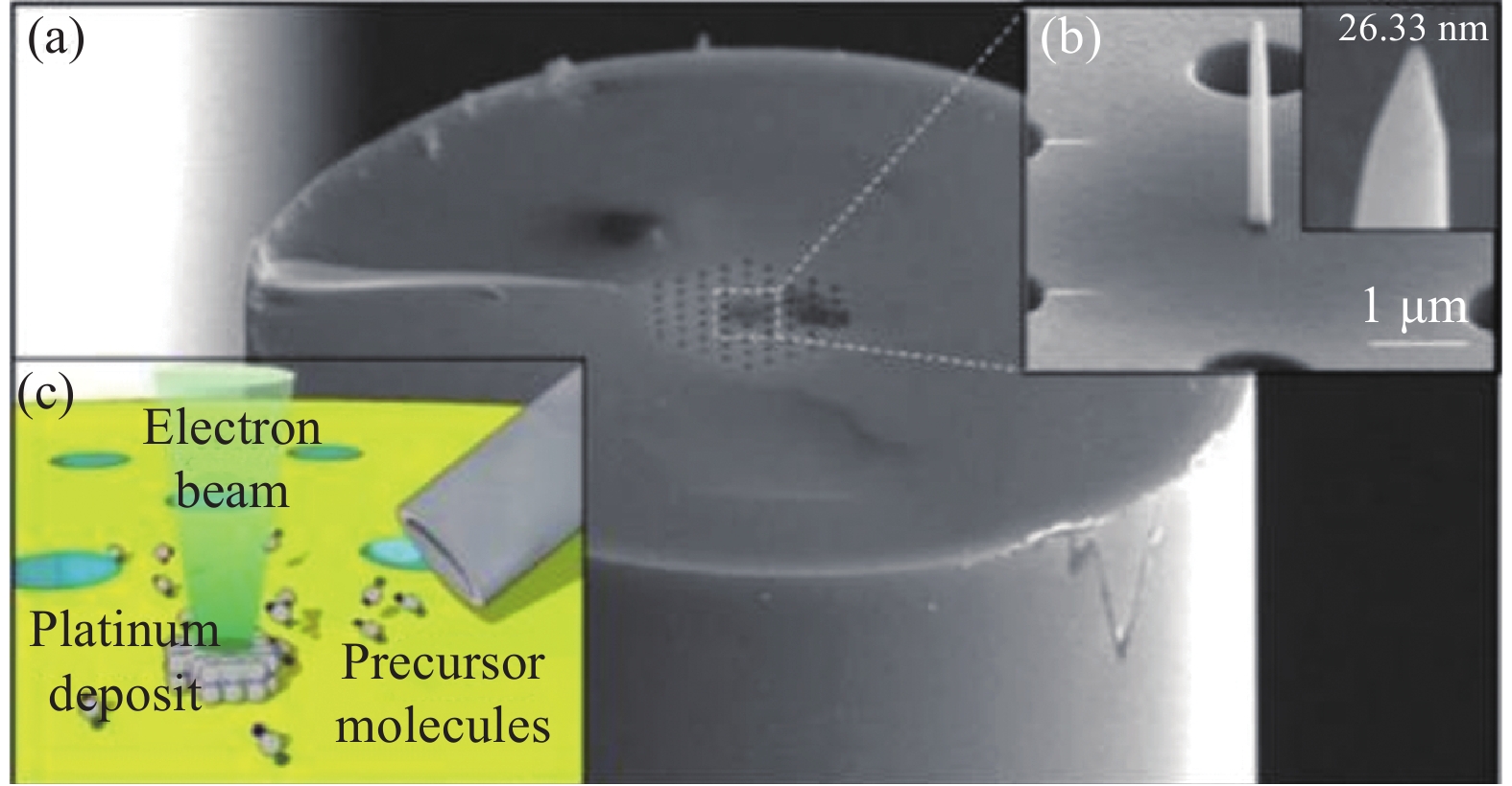
图 6 光纤光子晶体结合纳米天线
注:(a)等离激元天线结构;(b)纳米天线细节图;(c)诱导沉积过程图。
Figure 6. Nano-antennas combined with fiber optic photonic crystals
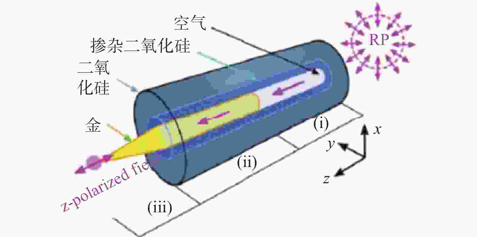
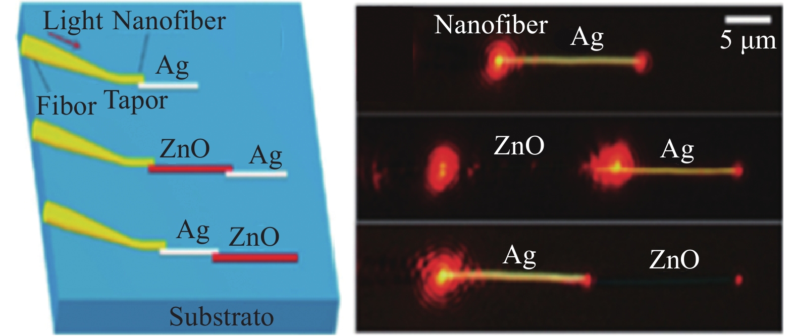
图 8 光纤包覆纳米线激发表面等离激元方法
Figure 8. Method of surface plasmon excitation using fiber coated with nanowires
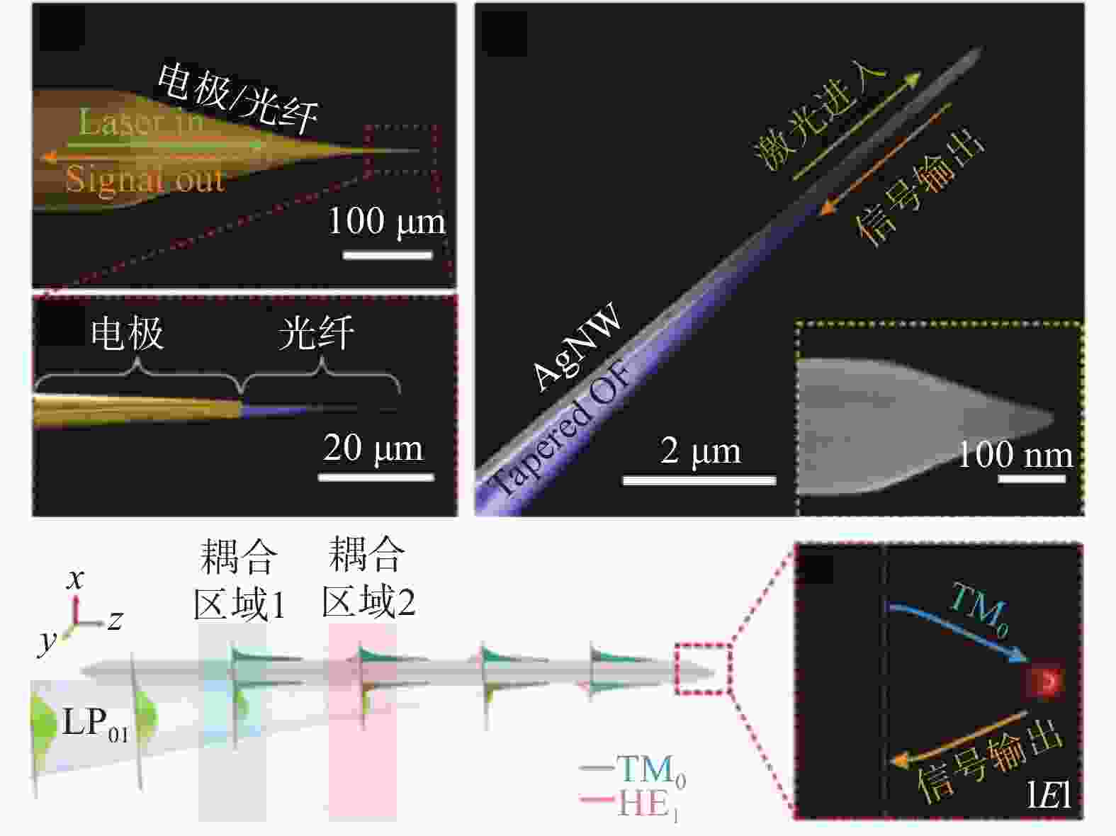
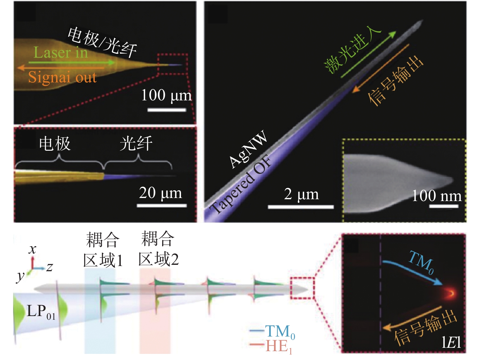
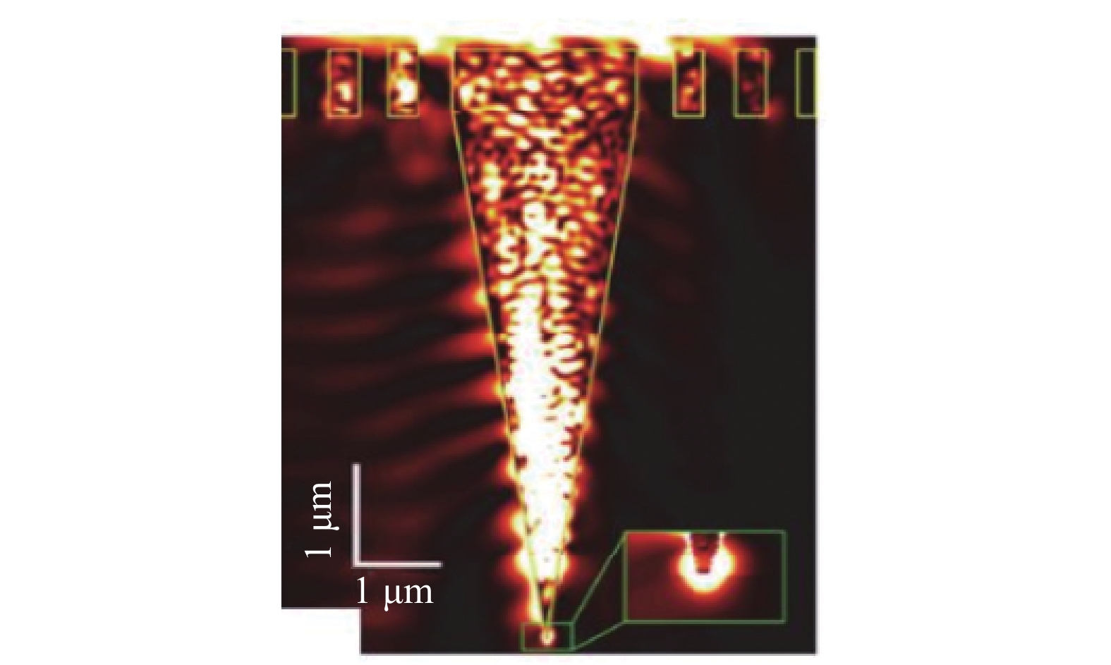
图 9 锥形光纤结合纳米线激发表面等离激元方法
Figure 9. Surface plasmon excitation method using conical fiber combined with nanowires
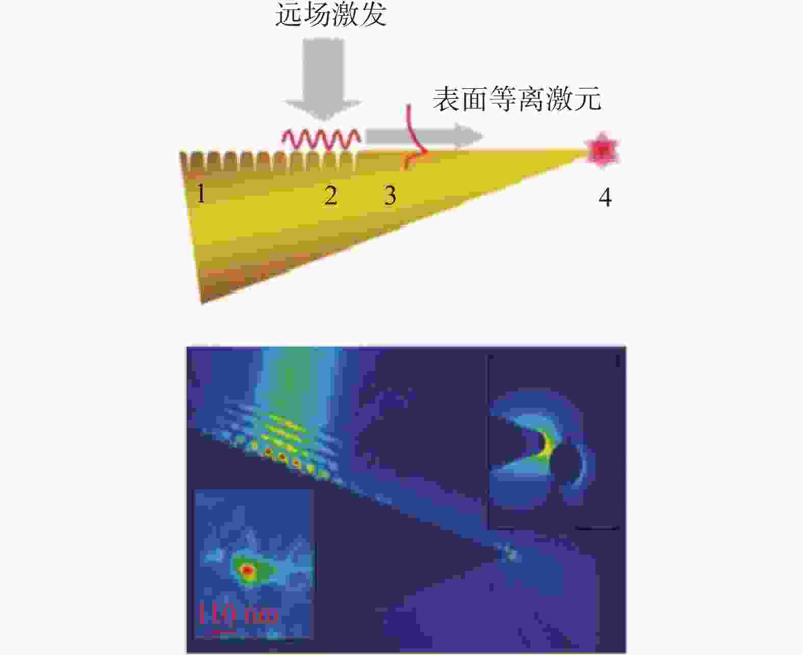
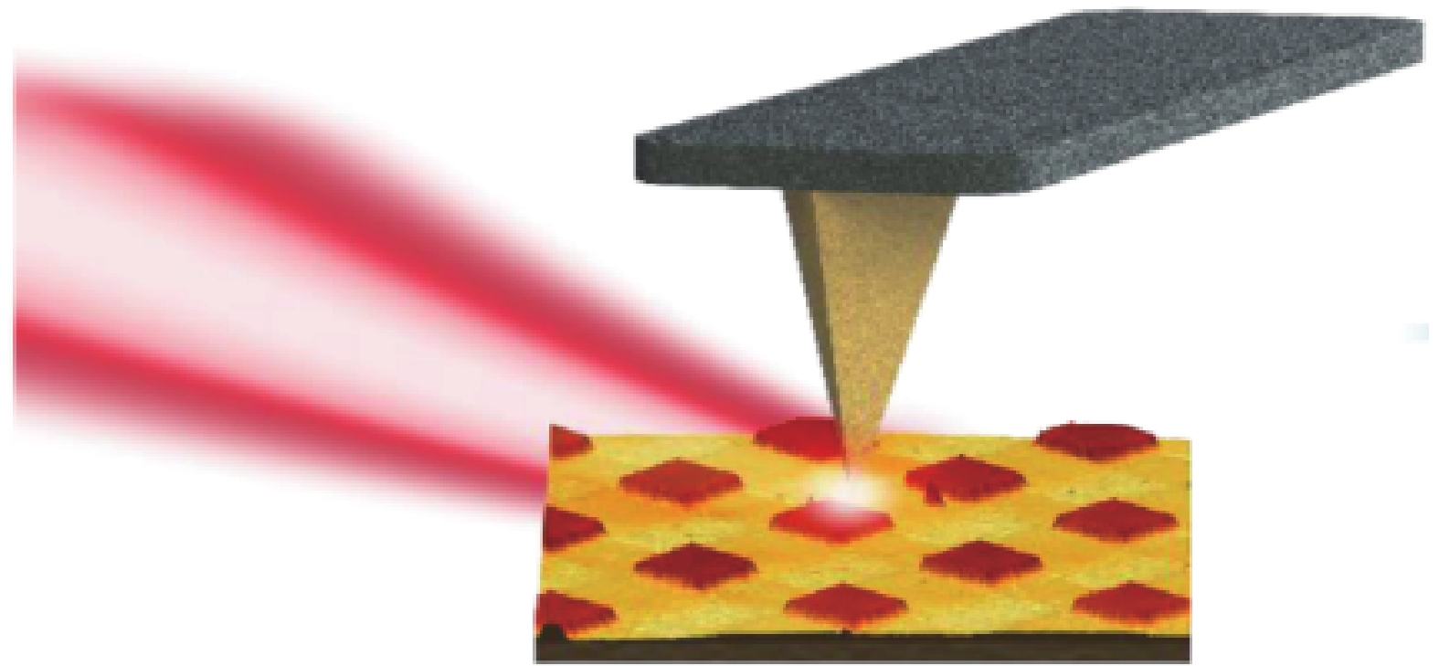
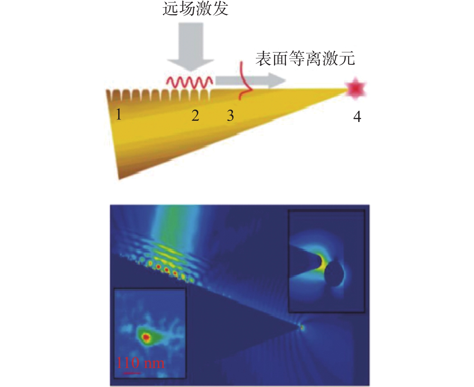
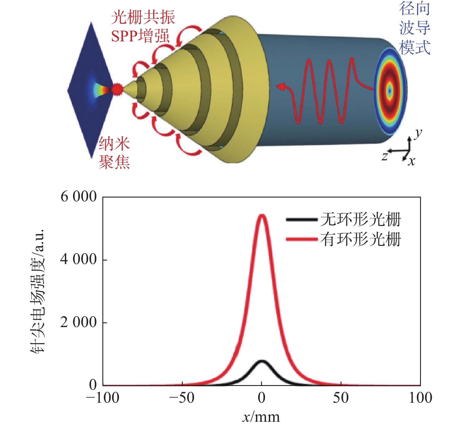
图 11 光栅激发表面等离激元示意图
Figure 11. Schematic of surface plasmon excitation by an optical grating
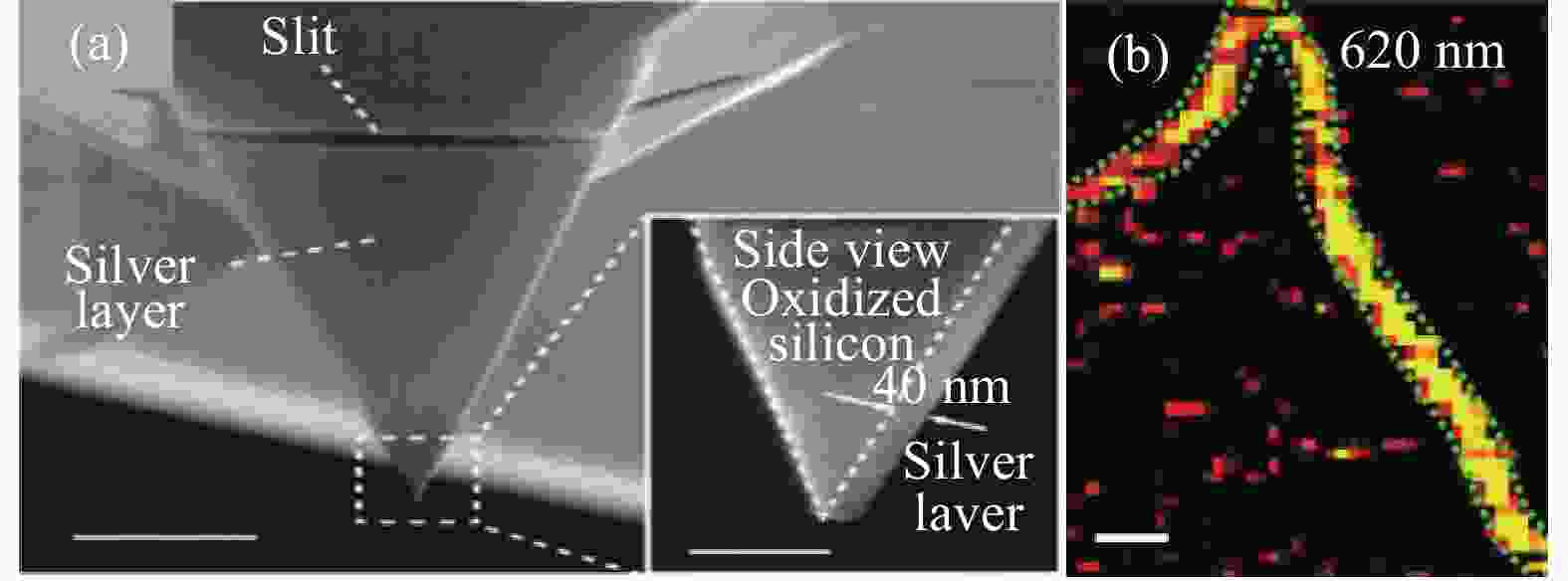
图 13 光栅耦合器及单壁碳纳米管测试结果
Figure 13. Test results of single-wall carbon nanotubes using a grating coupler
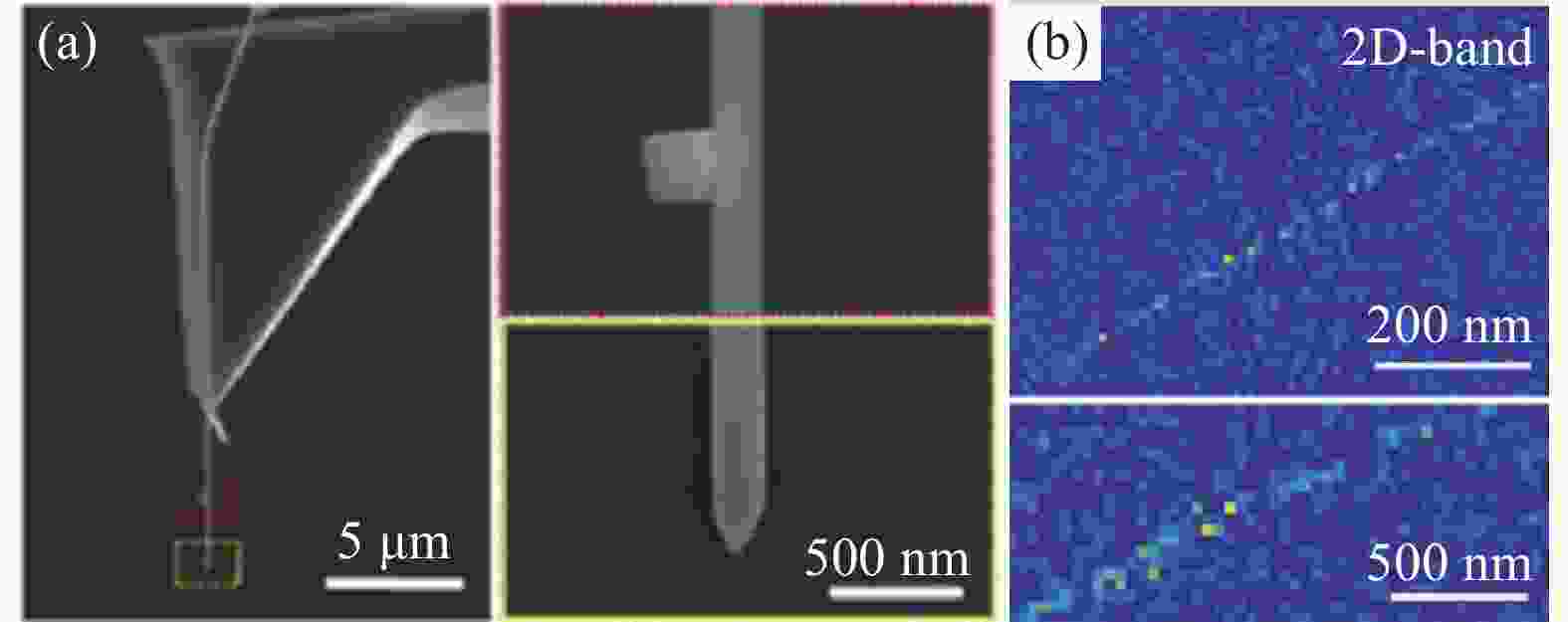
图 15 纳米线与纳米颗粒结构及测试结果
Figure 15. Nanowires and nanoparticle structures and test results

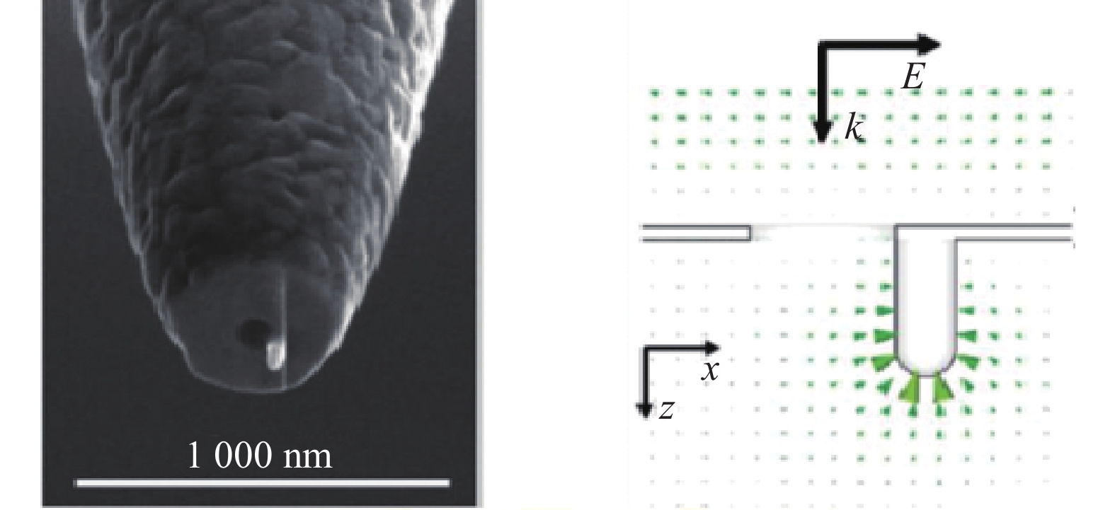
图 17 小孔衍射激发表面等离激元及聚焦过程
Figure 17. Surface plasmons excitation and focusing by aperture diffraction process
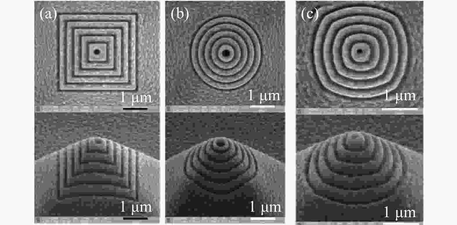
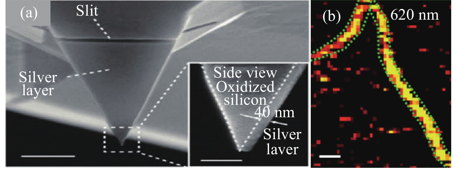
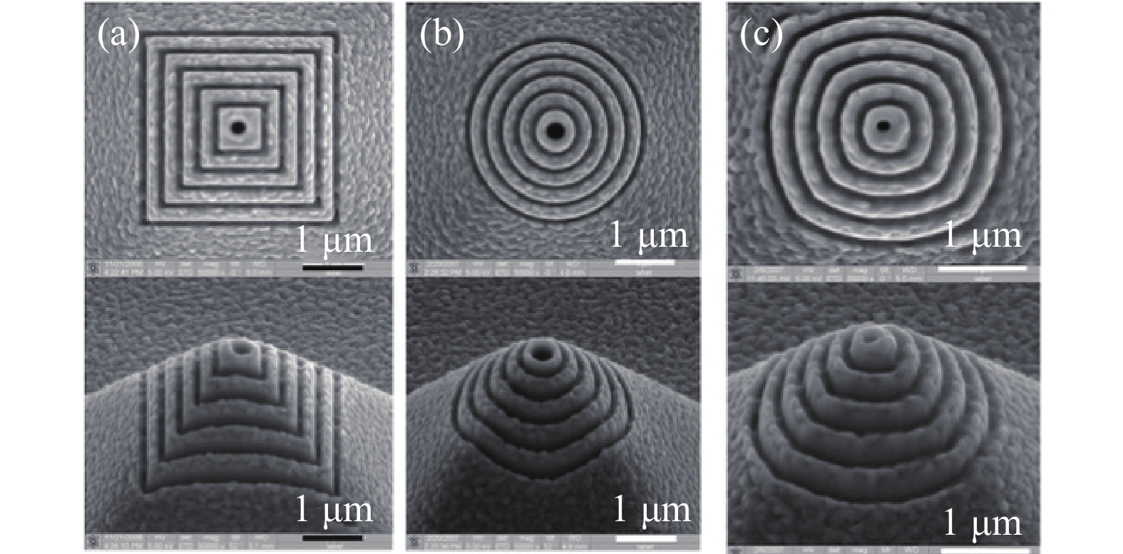
图 18 光纤探针尖端金字塔结构
注:(a)方形环;(b)圆形环;(c)椭圆环。
Figure 18. Pyramid structure at the tip of the optical fiber probe
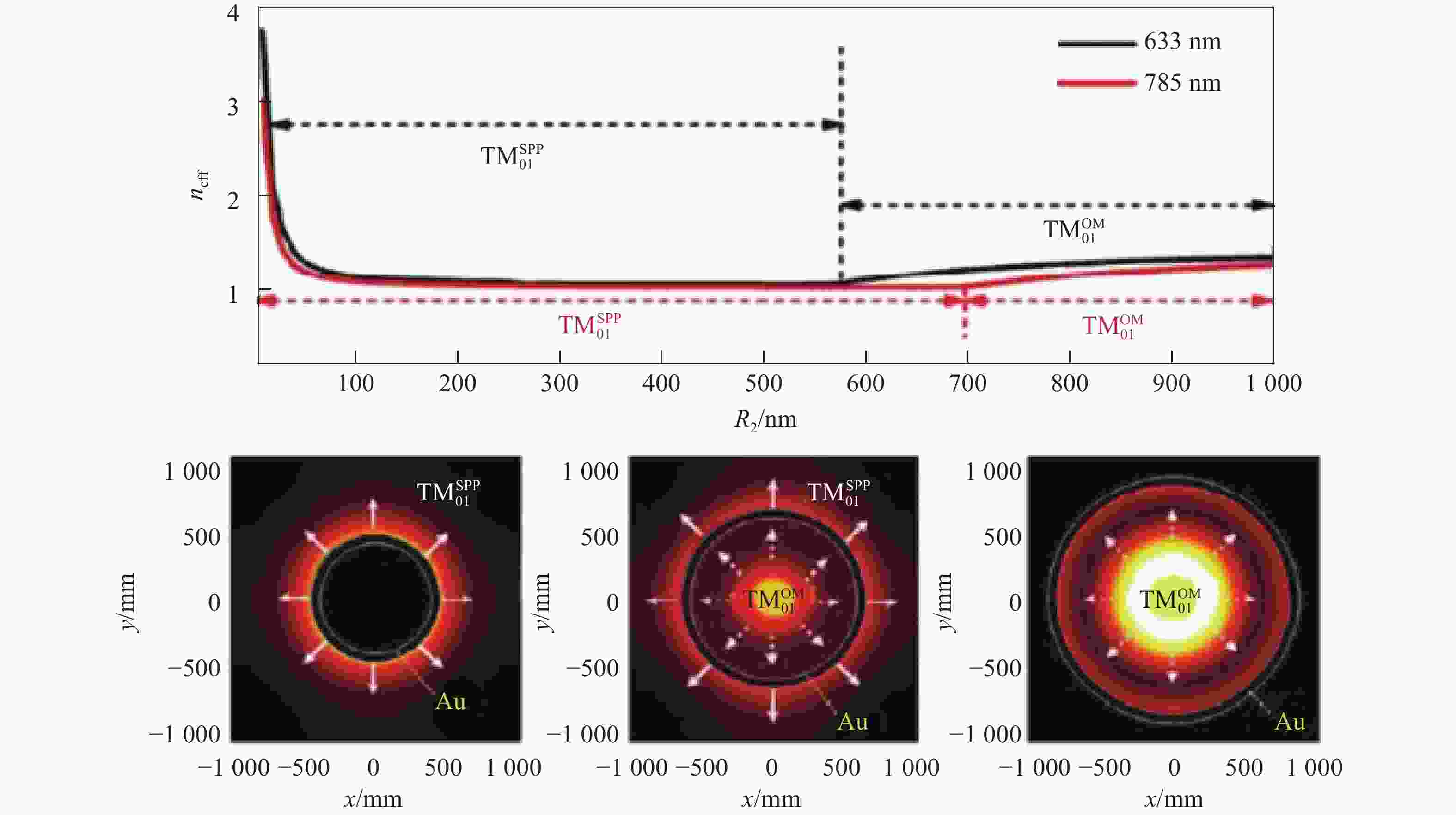
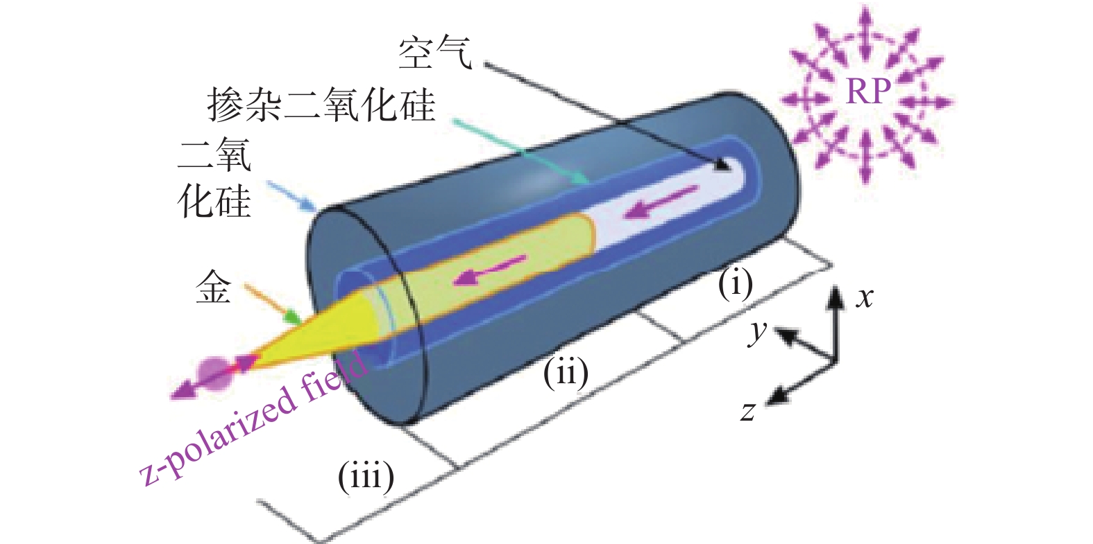
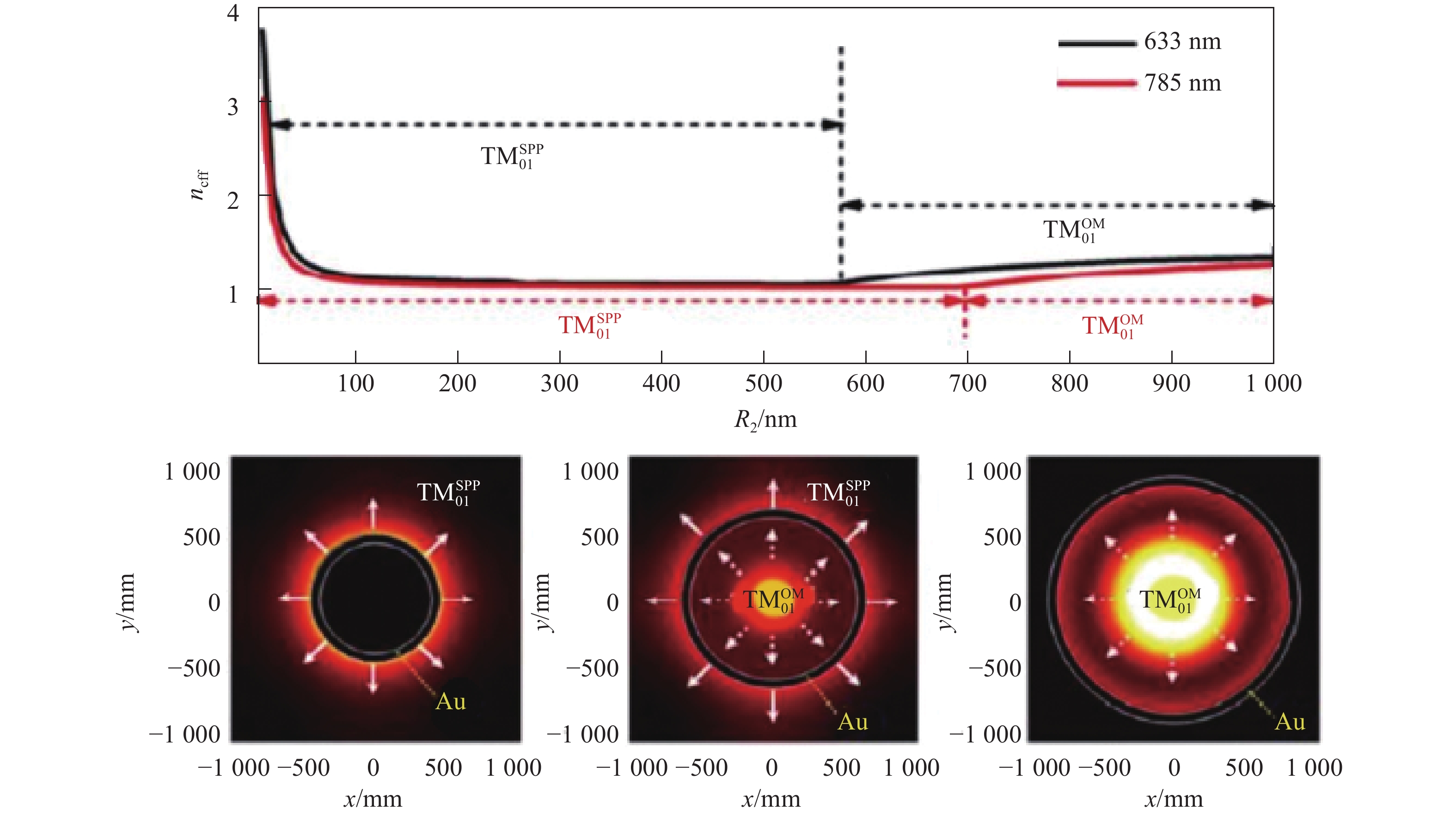
图 19 波导模式与等离激元模式耦合转化过程
Figure 19. Coupling and transformation process between waveguide and plasmon modes
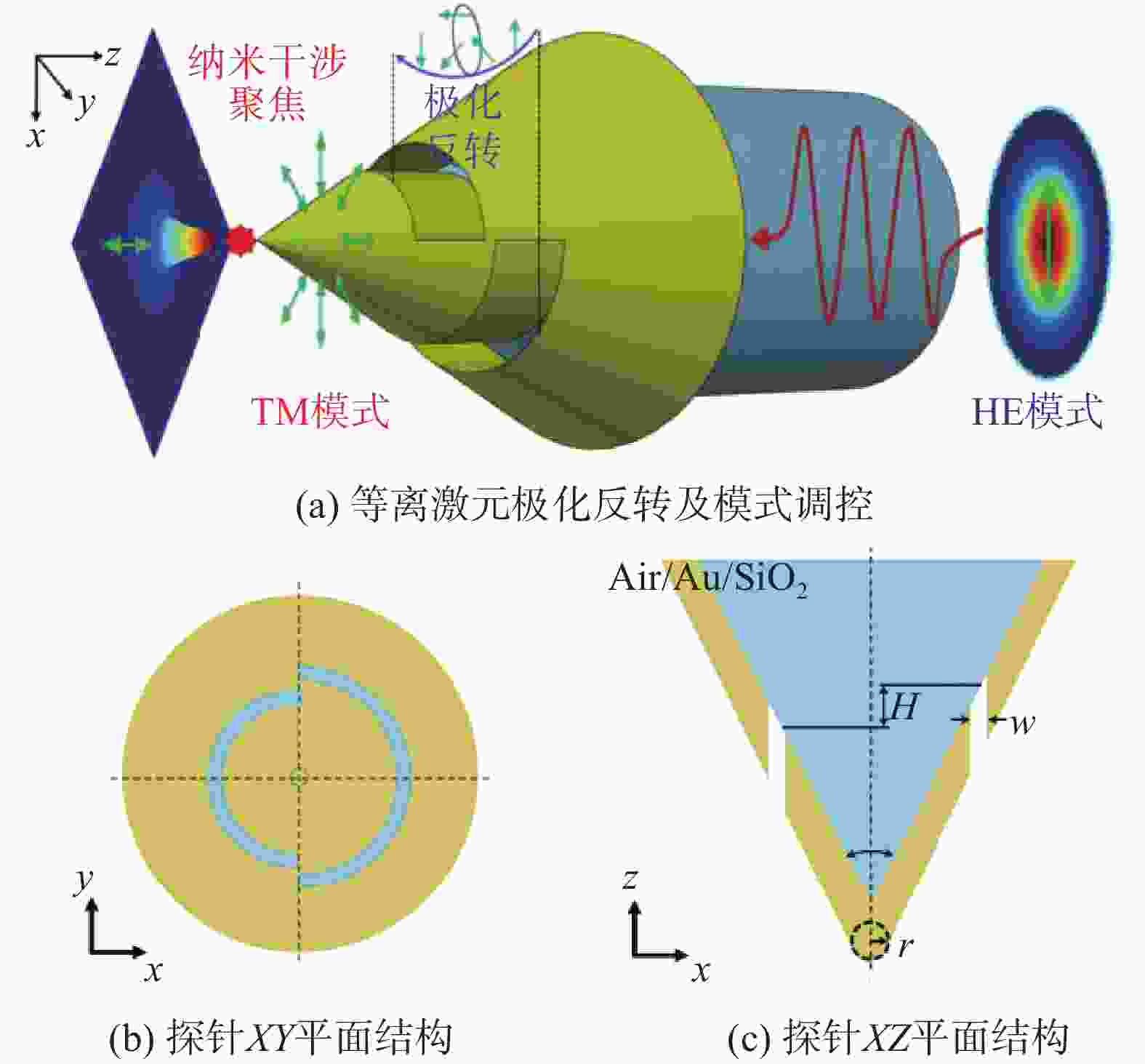
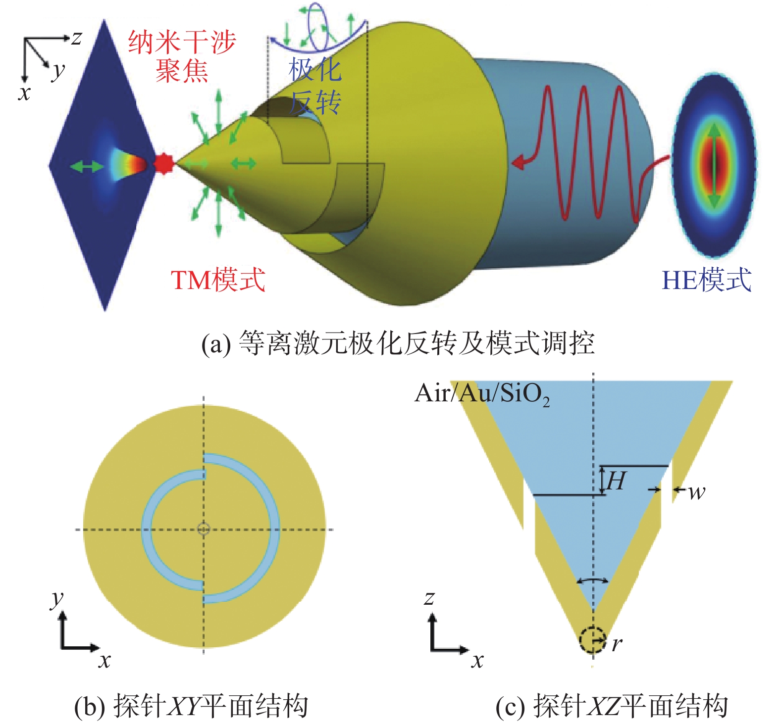
图 21 半环非对称离激元光纤探针及聚焦机理
Figure 21. Semi-ring asymmetric plasmon fiber probe and its focusing mechanism
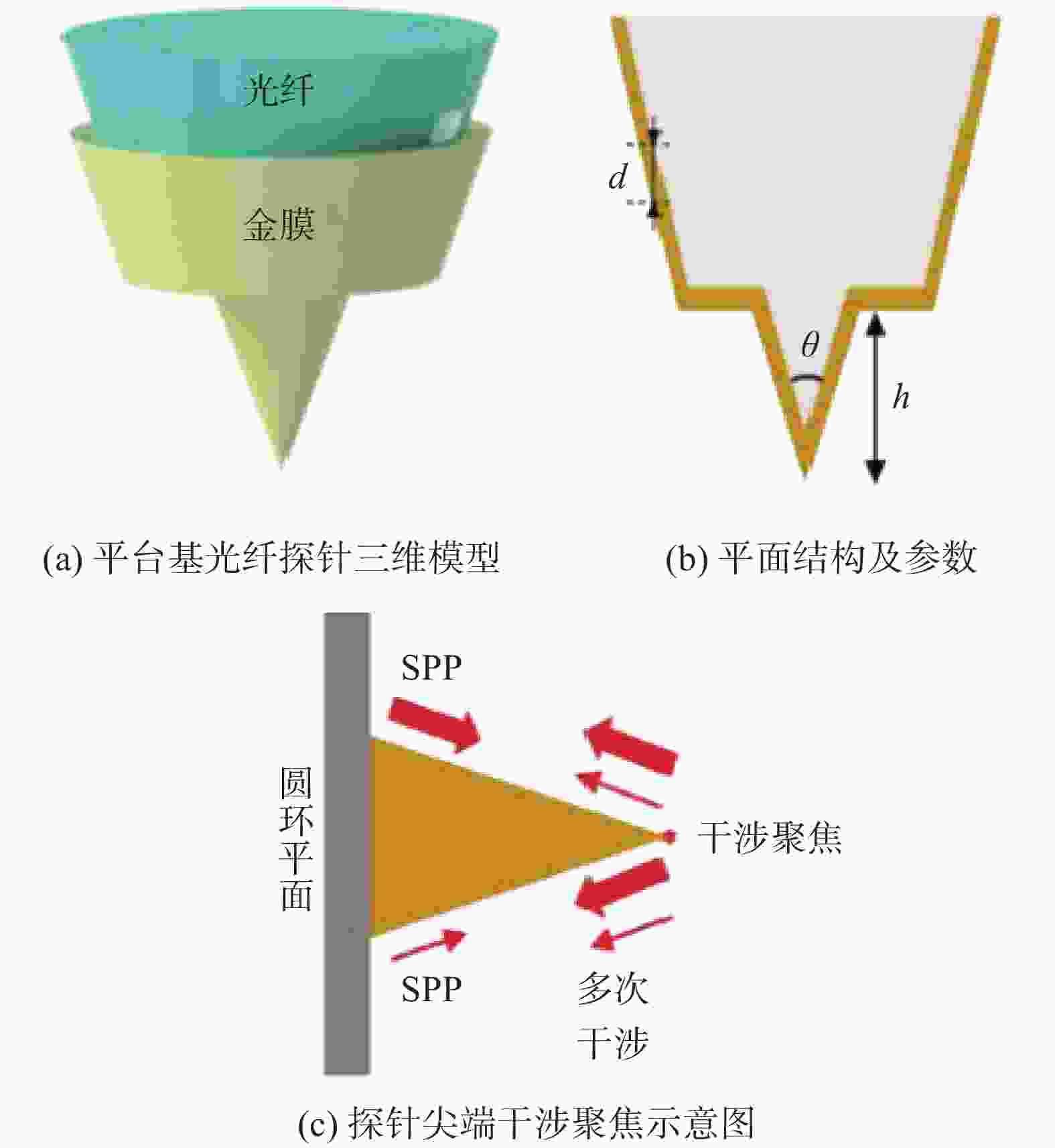
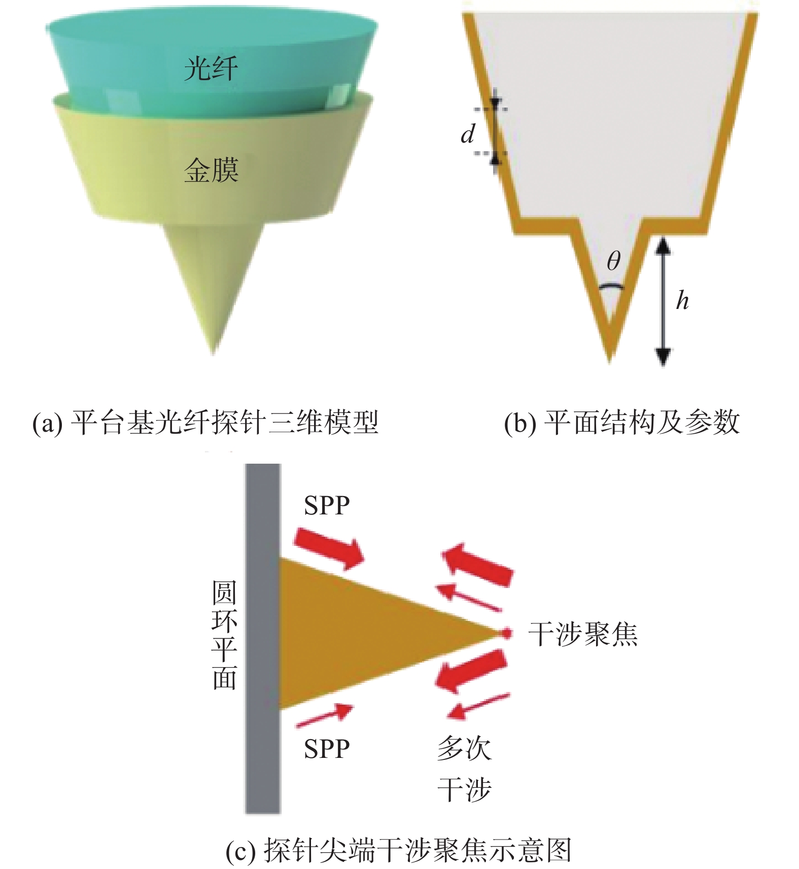
图 23 平台基光纤探针几何模型及干涉聚焦机理
Figure 23. Geometric model and interferometric focusing mechanism of a platform-based fiber probe
-
[1] LI Y, ZHAO F, CHENG X, et al. Four-Period Vertically Stacked SiGe/Si Channel FinFET Fabrication and Its Electrical Characteristics[J]. Nanomaterials, 2021, 11(7): 1689. doi: 10.3390/nano11071689 [2] ZHOU R, EDWARDS C, ARBABI A, et al. Detecting 20 nm Wide Defects in Large Area Nanopatterns Using Optical Interferometric Microscopy[J]. Nano Letters, 2013, 13(8): 3716-3721. doi: 10.1021/nl401622b [3] ZHONG H, LIU N, YANG Z, et al. Visible-Infrared Transparent Coding Metasurface Based on Random Metal Grid for Broadband Microwave Scattering[J]. Acs Applied Electronic Materials, 2021, 3(11): 4870-4876. doi: 10.1021/acsaelm.1c00699 [4] 艾俊强, 张扬, 王健. 电磁计算在飞机隐身设计中的应用及未来需求[J]. 电波科学学报, 2020, 35(1): 122-127. [5] 袁哲俊, 谢大纲, 胡忠辉. 微飞行器技术的最新发展[J]. 航空精密制造技术, 2005(1): 1-5. doi: 10.3969/j.issn.1003-5451.2005.01.001 [6] 陈耀文, 林月娟, 张海丹, 等. 扫描电子显微镜与原子力显微镜技术之比较[J]. 中国体视学与图像分析, 2006(1): 53-58. doi: 10.3969/j.issn.1007-1482.2006.01.013 [7] 凌妍, 钟娇丽, 唐晓山, 等. 扫描电子显微镜的工作原理及应用[J]. 山东化工, 2018, 47(9): 78-79,83. doi: 10.3969/j.issn.1008-021X.2018.09.033 [8] 刘剑霜, 谢锋, 吴晓京, 等. 扫描电子显微镜[J]. 上海计量测试, 2003(6): 37-39. doi: 10.3969/j.issn.1673-2235.2003.06.015 [9] 吴立新, 陈方玉. 现代扫描电镜的发展及其在材料科学中的应用[J]. 武钢技术, 2005(6): 36-40. doi: 10.3969/j.issn.1008-4371.2005.06.012 [10] 武开业. 扫描电子显微镜原理及特点[J]. 科技信息, 2010(29): 107. doi: 10.3969/j.issn.1001-9960.2010.29.080 [11] 朱琳. 扫描电子显微镜及其在材料科学中的应用[J]. 吉林化工学院学报, 2007(2): 81-84,92. doi: 10.3969/j.issn.1007-2853.2007.02.024 [12] 鲍幸峰, 方积年. 原子力显微镜在生物大分子结构研究中的应用进展[J]. 分析化学, 2000(10): 1300-1307. doi: 10.3321/j.issn:0253-3820.2000.10.028 [13] 刘小虹, 颜肖慈, 罗明道, 等. 原子力显微镜及其应用[J]. 自然杂志, 2002(1): 36-40. doi: 10.3969/j.issn.0253-9608.2002.01.007 [14] 张德添, 何昆, 张飒, 等. 原子力显微镜发展近况及其应用[J]. 现代仪器, 2002(3): 6-9. [15] 朱杰, 孙润广. 原子力显微镜的基本原理及其方法学研究[J]. 生命科学仪器, 2005(1): 22-26. doi: 10.3969/j.issn.1671-7929.2005.01.006 [16] 万瑾, 黄元庆. 激光三角法测量的研究[J]. 三明学院学报, 2006(4): 361-364. doi: 10.3969/j.issn.1673-4343.2006.04.001 [17] 郑德华, 沈云中, 刘春. 三维激光扫描仪及其测量误差影响因素分析[J]. 测绘工程, 2005(2): 32-34,56. doi: 10.3969/j.issn.1006-7949.2005.02.010 [18] ASH E A, NICHOLLS G. SUPER-RESOLUTION APERTURE SCANNING MICROSCOPE[J]. Nature, 1972, 237(5357): 510. doi: 10.1038/237510a0 [19] ZENHAUSERN F, MARTIN Y, WICKRAMASINGHE H K. SCANNING INTERFEROMETRIC APERTURELESS MICROSCOPY - OPTICAL IMAGING AT 10 ANGSTROM RESOLUTION[J]. Science, 1995, 269(5227): 1083-1085. doi: 10.1126/science.269.5227.1083 [20] HAROOTUNIAN A, BETZIG E, ISAACSON M, et al. SUPERRESOLUTION FLUORESCENCE NEAR-FIELD SCANNING OPTICAL MICROSCOPY[J]. Applied Physics Letters, 1986, 49(11): 674-676. doi: 10.1063/1.97565 [21] ISAACSON M, CLINE J, BARSHATZKY H. Near-field-optical microscopy[J]. AIP Conference Proceedings, 1991(241): 23-36. [22] ISAACSON M, CLINE J, BARSHATZKY H. RESOLUTION IN NEAR-FIELD OPTICAL MICROSCOPY[J]. Ultramicroscopy, 1992, 47(1-3): 15-22. doi: 10.1016/0304-3991(92)90182-J [23] SPECHT M, PEDARNIG J D, HECKL W M, et al. SCANNING PLASMON NEAR-FIELD MICROSCOPE[J]. Physical Review Letters, 1992, 68(4): 476-479. doi: 10.1103/PhysRevLett.68.476 [24] LIN J, MUELLER J P B, WANG Q, et al. Polarization-Controlled Tunable Directional Coupling of Surface Plasmon Polaritons[J]. Science, 2013, 340(6130): 331-334. doi: 10.1126/science.1233746 [25] RUCKSTUHL T, VERDES D, WINTERFLOOD C M, et al. Simultaneous near-field and far-field fluorescence microscopy of single molecules[J]. Optics Express, 2011, 19(7): 6836-6844. doi: 10.1364/OE.19.006836 [26] GRIGG D A, RUSSELL P E, GRIFFITH J E, et al. PROBE CHARACTERIZATION FOR SCANNING PROBE METROLOGY[J]. Ultramicroscopy, 1992, 42: 1616-1620. [27] MODY C C M. STARS: SCANNING PROBE MICROSCOPY[J]. Proceedings of the Ieee, 2014, 102(7): 1107-1112. doi: 10.1109/JPROC.2014.2326811 [28] REKHVIASHVILI S S. Modern methods of scanning-probe microscopy and spectroscopy[J]. Instruments and Experimental Techniques, 2002, 45(5): 724-726. doi: 10.1023/A:1020486925298 [29] TRANCA D E, STANCIU S G, HRISTU R, et al. High-resolution quantitative determination of dielectric function by using scattering scanning near-field optical microscopy[J]. Scientific Reports, 2015, 5: 11876. doi: 10.1038/srep11876 [30] TAUBNER T, KOROBKIN D, URZHUMOV Y, et al. Near-field microscopy through a SiC superlens[J]. Science, 2006, 313(5793): 1595. doi: 10.1126/science.1131025 [31] COURJON D, BAINIER C. NEAR-FIELD MICROSCOPY AND NEAR-FIELD OPTICS[J]. Reports on Progress in Physics, 1994, 57(10): 989-1028. doi: 10.1088/0034-4885/57/10/002 [32] GRAMOTNEV D K, BOZHEVOLNYI S I. Plasmonics beyond the diffraction limit[J]. Nature Photonics, 2010, 4(2): 83-91. doi: 10.1038/nphoton.2009.282 [33] REDDICK R C, WARMACK R J, FERRELL T L. NEW FORM OF SCANNING OPTICAL MICROSCOPY[J]. Physical Review B, 1989, 39(1): 767-770. doi: 10.1103/PhysRevB.39.767 [34] 邓燕. 浅谈倏逝波 [J]. 科技资讯, 2010 (13): 100, 3. [35] 胡三珍. 全内反射和倏逝波[J]. 华中师范大学学报(自然科学版), 1996(2): 169-173. [36] 李林, 肖循. 光的全反射中倏逝波的研究[J]. 武汉科技学院学报, 2006(12): 37-39. [37] 姚鸣. 全反射和倏逝波[J]. 宁夏工学院学报, 1995(4): 97-100. [38] 竺庆娥. 全反射时的倏逝波[J]. 北京师范大学学报(自然科学版), 1985(2): 82-85. [39] 陈龙, 何赛灵, 沈林放. 含负折射率介质的多层结构中倏逝波传播及隧道效应的分析[J]. 物理学报, 2003(10): 2386-2392. doi: 10.3321/j.issn:1000-3290.2003.10.006 [40] 黄惠杰, 翟俊辉, 任冰强, 等. 光纤倏逝波生物传感器及其应用[J]. 光学学报, 2003(4): 451-454. doi: 10.3321/j.issn:0253-2239.2003.04.015 [41] 倪旭, 张小柳, 卢明辉, 等. 声子晶体和声学超构材料[J]. 物理, 2012, 41(10): 655-662. [42] BACHELOT R, LERONDEL G, BLAIZE S, et al. Probing photonic and optoelectronic structures by apertureless scanning near-field optical microscopy[J]. Microscopy Research and Technique, 2004, 64(5-6): 441-452. doi: 10.1002/jemt.20102 [43] CADBY A, DEAN R, FOX A M, et al. Mapping the fluorescence decay lifetime of a conjugated polymer in a phase-separated blend using a scanning near-field optical microscope[J]. Nano Letters, 2005, 5(11): 2232-2237. doi: 10.1021/nl051525y [44] SYNGE E H. A suggested method for extending microscopic resolution into the ultra-microscopic region[J]. Philosophical Magazine, 1928, 6(35): 356-362. [45] GARCIA-PARAJO M F, VEERMAN J A, VAN NOORT S J T, et al. Near-field optical microscopy for DNA studies at the single molecular level[J]. Bioimaging, 1998, 6(1): 43-53. doi: 10.1002/1361-6374(199803)6:1<43::AID-BIO6>3.0.CO;2-F [46] IWABUCHI S, HASHIGASAKO A, MORITA Y, et al. Advanced imaging for DNA analysis based on scanning near-field optical/atomic-force microscopy (SNOAM) [C]. San Jose : Proceedings of the Conference on Scanning and Force Microscopies for Biomedical Applications, 1999. [47] JONGMIN K, OHTANI T, MURAMATSU H. Single molecule fluorescence imaging for its application to single DNA imaging by scanning near-field optical/atomic force microscopy [C]. 7th International Conference on Nanometer-Scale Science and Technology and 21st European Conference on Surface Science, 2002. [48] EL-KHOURY P Z. Tip-Enhanced Raman Scattering on Both Sides of the Schro''dinger Equation[J]. Accounts of Chemical Research, 2021, 54(24): 4576-4583. doi: 10.1021/acs.accounts.1c00597 [49] ZHANG R, ZHANG Y, DONG Z C, et al. Chemical mapping of a single molecule by plasmon-enhanced Raman scattering[J]. Nature, 2013, 498(7452): 82-86. doi: 10.1038/nature12151 [50] PAULITE M, BLUM C, SCHMID T, et al. Full Spectroscopic Tip-Enhanced Raman Imaging of Single Nanotapes Formed from β-Amyloid(1-40) Peptide Fragments[J]. Acs Nano, 2013, 7(2): 911-920. doi: 10.1021/nn305677k [51] LEE C, JEONG B G, KIM S H, et al. Investigating heterogeneous defects in single-crystalline WS2 via tip-enhanced Raman spectroscopy[J]. Npj 2d Materials and Applications, 2022, 6(1): 1. doi: 10.1038/s41699-021-00282-5 [52] HEINZELMANN H, POHL D W. SCANNING NEAR-FIELD OPTICAL MICROSCOPY[J]. Applied Physics a-Materials Science & Processing, 1994, 59(2): 89-101. [53] WANG H, WANG L, JAKOB D S, et al. Tomographic and multimodal scattering-type scanning near-field optical microscopy with peak force tapping mode[J]. Nature Communications, 2018, 9(1): 2005. doi: 10.1038/s41467-018-04403-5 [54] WANG H, WANG L, XU X G. Scattering-type scanning near-field optical microscopy with low-repetition-rate pulsed light source through phase-domain sampling[J]. Nature Communications, 2016, 7: 13212. doi: 10.1038/ncomms13212 [55] ANTOSIEWICZ T J, WROBEL P, SZOPLIK T. High resolution SNOM probes[C]. Polanica Zdroj: Proceedings of the 16th Polish-Slovak-Czech Optical Conference on Wave and Quantum Aspects of Contemporary Optics, 2008. [56] 胡德波, 戴庆. 低维纳米材料的近场光学表征[J]. 科学通报, 2018, 63(35): 3747-3759. [57] NOVOTNY L. From near-field optics to optical antennas[J]. Physics Today, 2011, 64(7): 47-52. doi: 10.1063/PT.3.1167 [58] MONTICONE F, ALU A. Metamaterials and plasmonics: From nanoparticles to nanoantenna arrays, metasurfaces, and metamaterials[J]. Chinese Physics B, 2014, 23(4): 12. [59] TAMINIAU T H, MOERLAND R J, SEGERINK F B, et al. lambda/4 Resonance of an optical monopole antenna probed by single molecule fluorescence[J]. Nano Letters, 2007, 7(1): 28-33. doi: 10.1021/nl061726h [60] MINN K, BIRMINGHAM B, KO B, et al. Interfacing photonic crystal fiber with a metallic nanoantenna for enhanced light nanofocusing[J]. Photonics Research, 2021, 9(2): 252-258. doi: 10.1364/PRJ.411583 [61] 高开, 唐浩, 栾中岳, 等. 表面等离激元应用研究新进展[J]. 化工新型材料, 2013, 41(1): 5-8. doi: 10.3969/j.issn.1006-3536.2013.01.002 [62] 童廉明, 徐红星. 表面等离激元——机理、应用与展望[J]. 物理, 2012, 41(9): 582-588. [63] 任升, 刘丽炜, 李金华, 等. 纳米尺度下的局域场增强研究进展[J]. 中国光学, 2018, 11(1): 31-46. [64] 黄锋. 基于纳米天线阵列的表面等离激元定向激发和聚焦 [D]. 哈尔滨: 哈尔滨工业大学, 2017. [65] HUANG J, YANG Z, WEI D, et al. Enhancement Effects of the Terahertz Near-Field Microscopy[J]. Applied Sciences-Basel, 2015, 5(4): 1745-1755. doi: 10.3390/app5041745 [66] KRUG J T, SáNCHEZ E J, XIE X S. Design of near-field optical probes with optimal field enhancement by finite difference time domain electromagnetic simulation[J]. Journal of Chemical Physics, 2002, 116(24): 10895-10901. doi: 10.1063/1.1479723 [67] LU F, ZHANG W, LIU M, et al. Tip-Based Plasmonic Nanofocusing: Vector Field Engineering and Background Elimination[J]. Ieee Journal of Selected Topics in Quantum Electronics, 2021, 27(1): 1. [68] MINN K, BIRMINGHAM B, ZHANG Z. New development of nanoscale spectroscopy using scanning probe microscope[J]. Journal of Vacuum Science & Technology A, 2020, 38(3): 38. [69] PIENPINIJTHAM P, KITAHAMA Y, OZAKI Y. Progress of tip-enhanced Raman scattering for the last two decades and its challenges in very recent years[J]. Nanoscale, 2022, 14(14): 5265-5288. doi: 10.1039/D2NR00274D [70] JING J, LIU K, JIANG J, et al. Performance improvement approaches for optical fiber SPR sensors and their sensing applications[J]. Photonics Research, 2022, 10(1): 126-147. doi: 10.1364/PRJ.439861 [71] 杨天, 陈成, 王晓丹, 等. 光纤端的等离激元探测技术[J]. 激光与光电子学进展, 2019, 56(20): 49-66. [72] 吴赟琨. 新型表面等离激元探针在量子光学中的应用 [D]. 合肥: 中国科学技术大学, 2021. [73] TUGCHIN B N, JANUNTS N, KLEIN A E, et al. Plasmonic Tip Based on Excitation of Radially Polarized Conical Surface Plasmon Polariton for Detecting Longitudinal and Transversal Fields[J]. Acs Photonics, 2015, 2(10): 1468-1475. doi: 10.1021/acsphotonics.5b00339 [74] TUNIZ A, SCHMIDT M A. Broadband efficient directional coupling to short-range plasmons: towards hybrid fiber nanotips[J]. Optics Express, 2016, 24(7): 7507-7524. doi: 10.1364/OE.24.007507 [75] TUNIZ A, CHEMNITZ M, DELLITH J, et al. Hybrid-Mode-Assisted Long-Distance Excitation of Short-Range Surface Plasmons in a Nanotip-Enhanced Step-Index Fiber[J]. Nano Letters, 2017, 17(2): 631-637. doi: 10.1021/acs.nanolett.6b03373 [76] KIM S, YU N, MA X, et al. High external-efficiency nanofocusing for lens-free near-field optical nanoscopy[J]. Nature Photonics, 2019, 13(9): 636-643. doi: 10.1038/s41566-019-0456-9 [77] WANG F, YANG S, LI S, et al. High resolution and high signal-to-noise ratio imaging with near-field high-order optical signals[J]. Nano Research, 2022, 15(9): 8345-8350. doi: 10.1007/s12274-022-4422-3 [78] ROPERS C, NEACSU C C, ELSAESSER T, et al. Grating-coupling of surface plasmons onto metallic tips: A nanoconfined light source[J]. Nano Letters, 2007, 7(9): 2784-2788. doi: 10.1021/nl071340m [79] ZHANG Z, AHN P, DONG B, et al. Quantitative Imaging of Rapidly Decaying Evanescent Fields Using Plasmonic Near-Field Scanning Optical Microscopy[J]. Scientific Reports, 2013, 3(1): 2803. doi: 10.1038/srep02803 [80] UMAKOSHI T, SAITO Y, VERMA P. Highly efficient plasmonic tip design for plasmon nanofocusing in near-field optical microscopy[J]. Nanoscale, 2016, 8(10): 5634-5640. doi: 10.1039/C5NR08548A [81] UMAKOSHI T, TANAKA M, SAITO Y, et al. White nanolight source for optical nanoimaging[J]. Science Advances, 2020, 6(23): 1. [82] FUJITA Y, WALKE P, DE FEYTER S, et al. Remote excitation-tip-enhanced Raman scattering microscopy using silver nanowire[J]. Japanese Journal of Applied Physics, 2016, 55(8): 3. [83] MA X, ZHU Y, YU N, et al. Toward High-Contrast Atomic Force Microscopy-Tip-Enhanced Raman Spectroscopy Imaging: Nanoantenna-Mediated Remote-Excitation on Sharp-Tip Silver Nanowire Probes[J]. Nano Letters, 2019, 19(1): 100-107. doi: 10.1021/acs.nanolett.8b03399 [84] GUO X, QIU M, BAO J, et al. Direct Coupling of Plasmonic and Photonic Nanowires for Hybrid Nanophotonic Components and Circuits[J]. Nano Letters, 2009, 9(12): 4515-4519. doi: 10.1021/nl902860d [85] HOU Y, MA C, WANG W, et al. A dual-use probe for nano-metric photoelectric characterization using a confined light field generated by photonic crystals in the cantilever[J]. Nano Research, 2021, 14(11): 3848-3853. doi: 10.1007/s12274-021-3304-4 [86] CHOI S S, PARK M J, HAN C H, et al. Fabrication of pyramidal probes with various periodic patterns and a single nanopore[J]. Journal of Vacuum Science & Technology B, 2015, 33(6): 1-7. [87] LU F, ZHANG W, ZHANG L, et al. Nanofocusing of Surface Plasmon Polaritons on Metal-Coated Fiber Tip Under Internal Excitation of Radial Vector Beam[J]. Plasmonics, 2019, 14(6): 1593-1599. doi: 10.1007/s11468-019-00951-8 [88] LI S, YANG S. Nanofocusing of a novel plasmonic fiber tip coupling with nanograting resonance[J]. Journal of Physics D-Applied Physics, 2020, 53(21): 8. [89] LI S, YANG S, WANG F, et al. Plasmonic interference modulation for broadband nanofocusing[J]. Nanophotonics, 2021, 10(16): 4113-4123. doi: 10.1515/nanoph-2021-0405 [90] Li S , Yang S . Nanofocusing of a novel plasmonic fiber tip coupling with nanograting resonance[J]. Journal of Physics D Applied Physics, 2020, 53: 21. [91] WANG F, LI S, ZHAO S, et al. A flat-based plasmonic fiber probe for nanoimaging[J]. Nano Research, 2023, 16(5): 7545-7549. doi: 10.1007/s12274-022-5297-z [92] SUN L, NI H, LIU X, et al. 3D Printed Asymmetric Nanoprobe for Plasmonic Nanofocusing under Internal Illumination[J]. Acs Photonics, 2018, 5(12): 4872-4879. doi: 10.1021/acsphotonics.8b01041 -

 下载:
下载:


 点击查看大图

计量
- 文章访问数: 296
- HTML全文浏览量: 87
- PDF下载量: 45
- 被引次数: 0

