作者投稿
作者投稿 专家审稿
专家审稿 编辑办公
编辑办公

Valuation Method for Milk Somatic Cell Count Reference Materials and the Uncertainty Evaluation
-
摘要: 建立了基于显微成像流式细胞技术对牛奶体细胞数量标准物质中的牛奶体细胞浓度进行测量的方法。该方法通过细胞核荧光染料溴化乙锭(Ethidium Bromide)对牛奶体细胞进行标记,显微成像流式细胞技术提供的单细胞多参数显微图像可有效辅助流式细胞分析方法中对目标细胞的圈门识别,从而提高圈门策略的正确性和重复性。通过多份不同体细胞浓度的牛奶体细胞标准物质样本的多次测量分析证明,该方法具有良好的室内重复性和日间重复性。建立了基于显微成像流式技术的牛奶体细胞数量标准物质中牛奶体细胞浓度测量方法的数学模型,分析确定了该测量方法不确定度主要来源于测量重复性、样本前处理、试剂加样、圈门操作和仪器测量体积偏差等。综合评定,针对研究制备的牛奶体细胞数量标准物质候选物V级(~1.0×10⁶ /mL),基于显微成像流式细胞技术的牛奶体细胞浓度测量方法的测量相对不确定度为4.84%。
-
关键词:
- 计量学 /
- 牛奶体细胞 /
- 细胞浓度 /
- 不确定度 /
- 显微成像流式细胞技术
Abstract: A method based on microscopic imaging flow cytometry was established to measure the concentration of milk somatic cells in the standard substance for milk somatic cell count. This method labels milk somatic cells using the fluorescent dye Ethium Bromide, and the single cell multi parameter microscopic images provided by flow cytometry can effectively assist in identifying target cells in flow cytometry analysis, thereby providing the correctness and repeatability of the gate strategy. Through multiple measurements and analysis of milk somatic cell standard substance samples with different concentrations of somatic cells, it has been proven that this method has good indoor and daytime repeatability. A mathematical model for measuring the concentration of milk somatic cells in a standard substance for milk somatic cell count based on microscopic imaging flow cytometry technology was established. The analysis determined that the uncertainty of this measurement method mainly comes from measurement repeatability, sample pretreatment, reagent addition, gate operation, and measurement volume. Comprehensive evaluation, targeting the candidate V-grade (~1.0×10⁶/mL) of the standard substance for milk somatic cell count prepared in the study, the relative uncertainty of the measurement method for milk somatic cell concentration based on microscopic imaging flow cytometry is 4.84%.-
Key words:
- metrology /
- milk somatic cells /
- cell concentration /
- uncertainty /
- microscopic imaging flow cytometry
-
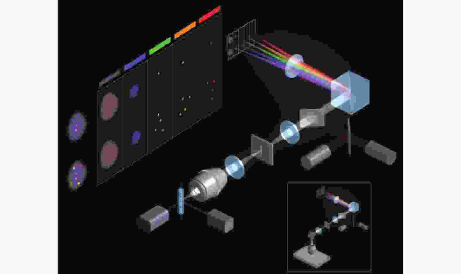
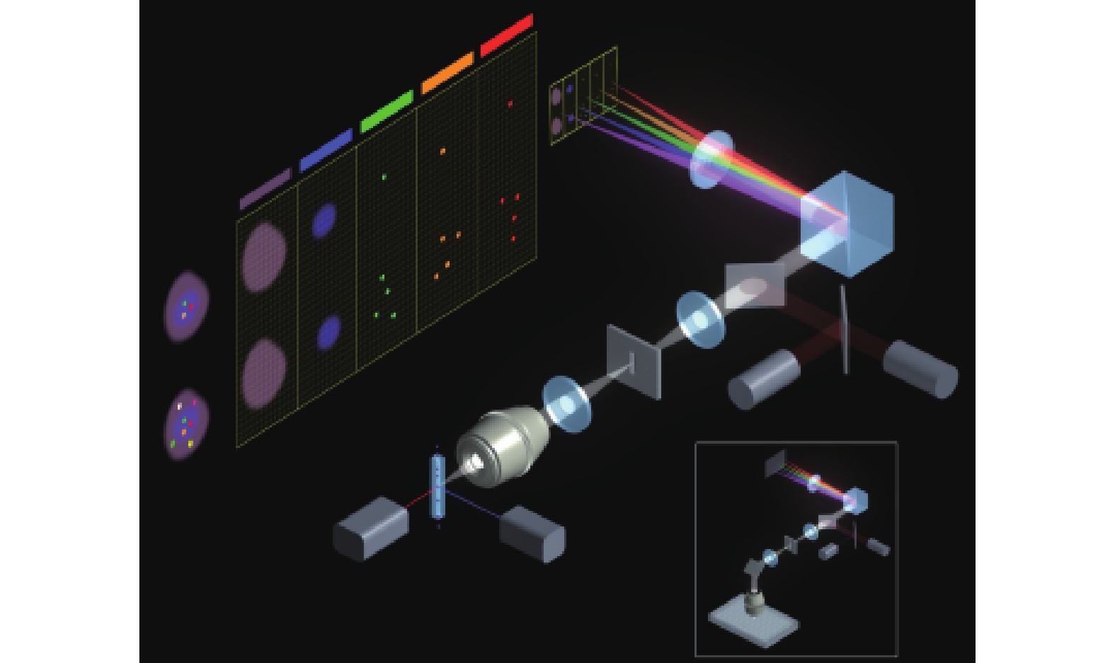
图 1 显微成像流式技术原理示意图
Figure 1. Schematic diagram of the principle of microscopic imaging flow cytometry technology
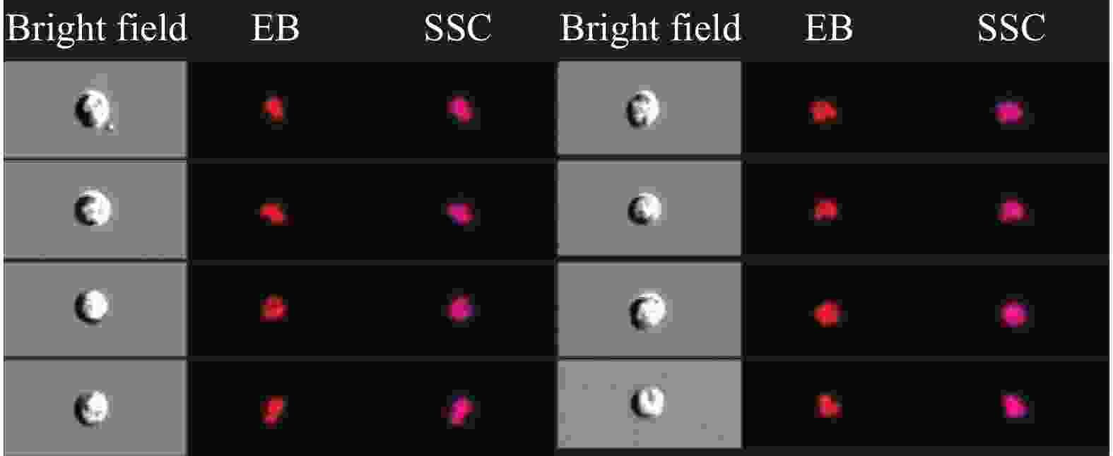
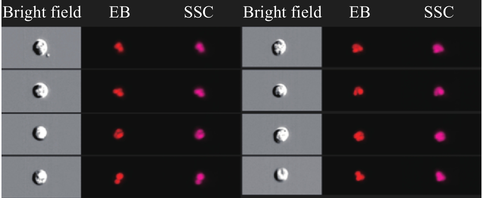
图 2 定值方法的流式细胞术圈门策略和单细胞显微3通道图像
Figure 2. Gating and multi-channel images of samples by microscopic imaging flow cytomety
表 1 测量方法室内重复性统计
Table 1. Indoor repeatability statistics of measurement methods
样本编号 1 2 3 4 5 6 7 I 细胞浓度(×106/m)L) 0.11 0.13 0.12 0.10 0.09 0.13 0.11 RSD(%) 3.6 3.1% 3.3% 2.9% 3.4% 3.7 3.7% II 细胞浓度(×106/m)L) 0.23 0.21 0.20 0.19 0.18 0.20 0.21 RSD(%) 2.7% 3.3% 3.0% 2.5% 3.1% 2.9% 2.7% III 细胞浓度(×106/m)L) 0.44 0.44 0.39 0.41 0.45 0.44 0.44 RSD(%) 2.9% 1.7% 2.0% 1.9% 2.6% 2.1% 2.6% IV 细胞浓度(×106/m)L) 0.90 0.88 0.80 0.85 0.89 0.83 0.88 RSD(%) 1.4% 1.7% 2.3% 2.3% 1.5% 1.9% 1.4% V 细胞浓度(×106/m)L) 1.06 1.05 1.08 1.05 0.99 0.98 1.01 RSD(%) 1.5% 1.2% 1.0% 1.4% 1.2% 1.4% 2.0%  下载: 导出CSV
表 2 测量方法日间重复性统计
Table 2. Measurement method daytime repeatability statistics
样本编号
时间(天)1 2 3 F P I 1 0.13±0.004 0.13±0.004 0.12±0.003 2.23 0.099 2 0.13±0.004 0.13±0.005 0.13±0.005 0.99 0.193 3 0.11±0.004 0.10±0.003 0.10±0.003 1.05 0.153 II 1 0.23±0.006 0.21±0.004 0.20±0.006 4.10 0.078 2 0.21±0.005 0.21±0.006 0.24±0.006 2.08 0.166 3 0.19±0.005 0.18±0.005 0.21±0.005 3.85 0.093 III 1 0.44±0.008 0.42±0.007 0.42±0.007 2.78 0.126 2 0.41±0.008 0.42±0.008 0.41±0.008 1.01 0.313 3 0.39±0.008 0.39±0.008 0.39±0.009 0.75 0.400 IV 1 0.90±0.013 0.90±0.014 0.90±0.013 0.95 0.359 2 0.88±0.015 0.89±0.015 0.89±0.014 1.89 0.0963 3 0.88±0.012 0.88±0.016 0.90±0.015 2.89 0.128 V 1 1.08±0.011 1.11±0.014 1.10±0.012 4.25 0.095 2 1.05±0.014 1.05±0.016 1.04±0.016 1.07 0.177 3 0.99±0.012 1.00±0.015 1.01±0.015 2.75 0.333
下载: 导出CSV
表 3 前处理方法细胞回收率数据分析
Table 3. Analysis of Cell Recovery Data for Pre processing Methods
样本编号 处理前(×106/mL) 处理后(×106/mL) 回收率(%) 1 1.02 0.99 96.6 2 1.10 1.03 93.6 3 0.96 0.91 94.5 4 0.99 0.94 95.0 5 1.07 0.99 92.3 6 1.12 1.02 90.8 7 1.07 0.98 91.6 8 0.92 0.86 93.3 9 0.99 0.95 95.6 10 1.13 1.07 94.6 平均回收率 93.8% RSD 1.95%
下载: 导出CSV
表 4 测量不确定度分量汇总表
Table 4. Summary of Measurement Uncertainty Components
不确定度来源 量值 标准不确定度u 相对不确定度urel(%) 测量重复性u1 10000 123.5 1.24 前处理细胞回收u2 93.8% 1.8% 1.95 试剂加样u3 0.83 0.024 0.015 圈门操作u4 10000events 50events 0.50 仪器测量体积偏差u5 100μL 0.5μL 0.50 测量相对不确定度 2.42% 测量扩展不确定度 4.84%
下载: 导出CSV
-
[1] 唐永明, 王锡波. 浅谈牛奶体细胞数在奶牛饲养管理中的应用[J]. 草食家畜, 2006(1): 47-48. [2] 汪悦, 王炳, 苏汉书, 等. 牛奶体细胞生成与乳品质量和安全的关系[J]. 动物营养学报, 2017, 29(7): 2269-2277. [3] 田树清, 范艳平. 乳体细胞数(SCC)在奶牛养殖业中的应用[J]. 中国奶牛, 2009(7): 46-47. [4] 刘晶. 牛奶体细胞数在牧场管理中的应用[J]. 中国乳业, 2012(5): 37-37. [5] 杨佳怡, 牛春艳, 刘瑛颖, 等. 生鲜牛乳体细胞检测与计量校准必要性研究[J]. 生物技术通报, 2020(5): 16-21. [6] 中国农垦乳业联盟. 中国农垦乳业联盟产品标准 生鲜乳: T/SFLA 001—2019[S]. 北京: 中国农垦经贸流通协会, 2019: 11. [7] 中华人民共和国农业行业标准. 生鲜牛乳中体细胞测量方法: NY/T800-2004[S]. 北京: 中华人民共和国农业部, 2004: 6. [8] Patelp M, Bhata, Markx G H, et al. A comparative study of cell death using electrical capacitance measurements and diele etrophoresis[J]. Enzyme Micmb Tech, 2008, 43(7): 523-530. doi: 10.1016/j.enzmictec.2008.09.006 [9] 马保臣, 秦卓明, 李建基, 等. 奶牛隐性乳腺炎病原菌的分离鉴定和体细胞数及酶的相关性研究[J]. 家畜生态学报, 2006, 27(2): 63-67. [10] 左仪. 几种体细胞定量测定方法的应用[J]. 中国乳品工业, 1995, 23(1): 36-39. [11] 方海田, 刘慧燕, 德力格尔桑, 等. 牛乳乳汁电导率变化与体细胞数相关关系的研究[J]. 现代食品科技, 2007, 23(4): 86-88. [12] 胡松华, 蔡渭明, 杜爱芳, 等. 牛乳电导率和体细胞含量及乳腺感染关系的研究[J]. 中国兽医科技, 1993, 23(6): 1l-13. [13] 张莹蕾, 汪银锋, 李素平. 牛奶体细胞数的研究进展[J]. 河南畜牧兽医(综合版), 2008, 29(12): 9-10. [14] Davis B. H. , Roussel M. , Fest T. , et al.Toward a reference method for leukocyte differential counts in blood: Comparison of three flow cytometric candidate methods[J]. Cytometry, Part A: the journal of the International Society for Analytical Cytology, 2012, 81A(11). [15] Heung Kyu Lee, Jennifer M. Lund, Balaji Ramanathan, et al.Autophagy-Dependent Viral Recognition by Plasmacytoid Dendritic Cells [J]. Science, 2007, 315(5817): 1398-1401. [16] Rees P , Summers H D , Filby A , et al. Imaging flow cytometry[J]. Nature Reviews Methods Primers, 2022, 2(1). DOI: 10.1038/s43586-022-00167-x. [17] Cerrato G , Liu P , Martins I , et al. Quantitative determination of phagocytosis by bone marrow-derived dendritic cells via imaging flow cytometry[J]. Methods in Enzymology, 2019, 2: 632. [18] User Manual: XPR & XSR Analytical Balances [19] 倪育才. 实用测量不确定度评定[M]. 北京: 中国计量出版社, 2009. [20] 孙乃峰. 浅谈DHI测试报告中的体细胞数(scc)[J]. 中国奶牛, 2008(4): 63-64. [21] 石璞, 许尚忠. 奶牛DHI中的体细胞测定与牧场管理[J]. 中国奶牛, 2007(6): 22-24. doi: 10.3969/j.issn.1004-4264.2007.06.010 [22] Milk-Enumeration of somatic cells-Part 1: Microscopic method (Reference method): ISO 13366-1: 2008. [S/OL].https://www.iso.org/standard/40259.html [23] 张文阁, 刘俊杰. 血细胞技术分析用国家标准物质的研制[J]. 中国粉体技术, 2009(16): 59-64. [24] BD Multitest™ 6-color TBNK reagent with optional BD Trucount™ tube. [EB/OL]. [2021-04-01].https://www.bdbiosciences.com/cn/reagents/clinical/systems-kits/bd-trucount-tubes/c/782111. -

 点击查看大图
计量
- 文章访问数: 74
- HTML全文浏览量: 45
- PDF下载量: 8
- 被引次数: 0